Use the enhanced 38-structure skeletal lab alongside the assigned readings and virtual investigations. A complete body, soft-tissue systems, and physiological simulation are outside this viewer’s scope.
Choose a station and inspect its spatial relationships. Pin up to three structures, use a ghost view, and compare a visual cutaway.
Complete a guided five-structure mission, then repeat in retrieval practice with names hidden. Use reveals as learning aids and record them honestly.
Write a source-based reflection, identify an uncertainty, and export your local practice record for faculty discussion. Practice does not award a grade or verified clinical hours.
A keyboard-accessible structure list provides the same selection and practice pathway. Desktop learning works without a headset. The clinical room uses synthetic learning prompts and is also embedded in ChartRight SimEHR.
Start here
Course syllabus
Study human organization from chemistry to cells, tissues, and organ systems. Connect anatomical relationships with homeostasis, integument, bone, joints, muscle, nervous pathways, special senses, and endocrine regulation. Apply the ideas in virtual investigations and fictional clinical cases, ending with a three-case evidence portfolio.
DBIO 201 carries 3 lecture credits and DBIO 201L carries 1 laboratory credit in the proposed adopting-institution course structure. The public Deneb sample awards no institutional credit or professional certification. An institution must approve credit, contact-hour equivalence, instructor assignment, assessment security, and applicable policies before credit-bearing use.
Prerequisites
No prerequisite is imposed by this public sample. Prior introductory biology and college-level reading are recommended. An adopting institution sets its own enrollment requirements. Terminology and optional text-first alternatives support learners with different starting points.
Pacing and workload
The suggested sequence spans 16 weeks, with an 8-week option that pairs consecutive weeks. The planning estimate is 135 hours of lecture-related learning and 45 hours of laboratory-related learning, including independent study, for 180 total hours. These are workload estimates, not a verified contact-hour or accreditation determination. In public self-paced mode, suggested dates never lock access.
Materials and technology
Required reading is the freely accessible OpenStax Anatomy and Physiology 2e web text. Original Deneb lessons, model datasets, attributed illustrations, and browser-based models are included. Use an internet-connected browser, a way to write or dictate notes, and a spreadsheet or paper for graphs. No paid textbook, Quizlet class, medical equipment, biological specimen, or patient record is required.
Participation and submission
Read objectives, complete the lesson, attempt its activity, compare the feedback, complete the module lab and case, then take the checkpoint. Keep an evidence notebook. In the public sample, notes and practice results remain in your browser; export them before clearing storage or changing devices. Lab reports and portfolios are drafted locally. There is no public assignment upload or faculty grade synchronization.
Assessment and feedback
Public auto-scored practice contains twelve 20-point checkpoints and three 120-point examinations, totaling 600 points. Three attempts are available and the best score is retained; feedback is shown after submission. A 70% browser practice goal is for study only. The proposed 1000-point institutional scheme adds 150 portfolio points and 250 lab points using the rubrics. Those manually reviewed items are not included in the public browser score.
Academic integrity and AI
Attempt questions and predictions independently before checking feedback. AI may support vocabulary, study planning, or revision if the adopting instructor allows it. Verify factual claims against assigned sources, acknowledge permitted AI assistance, and do not enter patient, student, or other private information. A public practice answer bank is unsuitable for a secure institutional examination without replacement or supervised controls.
Accessibility
Every included illustration has a text description and attribution. Activities support typed, dictated, or equivalent text responses. Models have labeled controls and written outputs; datasets are also supplied as text tables. Quizzes are untimed. No personal exercise, painful stimulus, sensory challenge, or self-examination is required. An adopting institution supplies its approved accommodation contact and equivalent access process.
Clinical scope
Clinical examples teach structure and function and use fictional observations. They do not diagnose or prescribe. Synthetic lab datasets and arbitrary model ranges are labeled at their point of use. Clinical and academic review is pending. The public sample does not certify hands-on laboratory or clinical competence.
Institutional policies
Before credit-bearing delivery, the adopting institution must supply instructor identity and office hours, accommodation and support contacts, course dates, attendance and late-work rules, withdrawal policy, grading scale, lab competency requirements, and appeal procedures. These decisions remain institution-specific rather than being represented as approved Deneb policies.
Course learning outcomes
Explain organization from chemical components to cells tissues organs and systems.
Apply anatomical position directional terms planes and skeletal landmarks.
Connect tissue and organ structure with a stated physiological function.
Trace negative feedback and explain limits of a regulated response.
Compare normal function with selected mechanisms of injury and disease without diagnosing a person.
Interpret modeled observations and calculations with units assumptions and uncertainty.
Analyze responses to a changed input across interacting body systems.
Identify representative tissues and anatomical structures from attributed illustrations and descriptions.
Construct an evidence-supported explanation of a fictional cross-system case.
Complete virtual investigations with predictions observations analysis and stated limitations.
Suggested 16 week sequence
Weeks
Module
Lecture study h
Lab study h
Outcomes
1
Foundations of anatomy and physiology
7
3
CLO 1, CLO 2, CLO 4, CLO 6
2
Chemistry and cellular energy
7
3
CLO 1, CLO 3, CLO 4, CLO 6
3
Cells membranes and the cell cycle
8
3
CLO 1, CLO 3, CLO 4, CLO 6
4
Histology injury and repair
8
4
CLO 3, CLO 5, CLO 6, CLO 8
5
Integumentary system
8
3
CLO 3, CLO 4, CLO 5, CLO 8
6 to 7
Bone and the skeleton
14
5
CLO 2, CLO 3, CLO 5, CLO 8
8
Joints and movement
8
3
CLO 2, CLO 3, CLO 5, CLO 6
9 to 10
Muscle tissue and movement
16
4
CLO 2, CLO 3, CLO 5, CLO 6
11 to 12
Nervous tissue and central peripheral and autonomic pathways
22
7
CLO 2, CLO 3, CLO 4, CLO 5, CLO 6, CLO 8
13
Special senses
10
3
CLO 2, CLO 3, CLO 5, CLO 6, CLO 8
14 to 15
Endocrine system
17
4
CLO 3, CLO 4, CLO 5, CLO 6, CLO 7
16
Human systems integration
10
3
CLO 3, CLO 4, CLO 5, CLO 6, CLO 7, CLO 9, CLO 10
Total planning workload: 135 lecture-related hours and 45 laboratory-related hours. Modules use chemistry before transport, histology before organs, and electrical signaling before neural pathways.
How this course is designed to help you learn
Anatomy and physiology combine a large vocabulary with cause-and-effect mechanisms. The added features give you repeated retrieval of structures and processes, diagrams of mechanisms that photographs cannot show, and practice explaining why a change occurs.
Each module now contains a consistent set of study features drawn from learning research. Use them in order; they take about 20 to 30 extra minutes per module and are not scored.
Feature
What you do
Why it helps
Module roadmap
Preview the sequence, time, and objectives.
Advance organizers and clear signaling reduce extraneous load and help you connect new ideas to a structure (Mayer, 2021).
Make a prediction
Answer one question before the lessons.
Pretesting improves later learning of the tested content, even after an error, if feedback follows (Richland et al., 2009; Pan & Carpenter, 2023).
Retrieval check after each lesson
Answer from memory, then read the explanation.
Practice testing is among the most effective study methods; explanatory feedback corrects errors (Roediger & Karpicke, 2006; Dunlosky et al., 2013; Hattie & Timperley, 2007).
New diagrams with text descriptions
Study the picture and its words together.
Words with related graphics support deeper understanding than words alone; text descriptions keep the content accessible (Mayer, 2021; CAST, 2024).
Watch for these errors
Compare a common error with better reasoning.
Refutation text that names and corrects a misconception changes understanding more than explanation alone (Tippett, 2010).
Self-explanation and elaborative questioning improve transfer (Chi et al., 1994; Dunlosky et al., 2013).
Spaced review
Answer questions from earlier modules.
Distributed and interleaved practice improves retention and method selection (Cepeda et al., 2006; Rohrer & Taylor, 2007).
Close the module
Free recall, confidence rating, muddiest point.
Self-monitoring and calibration support self-regulated learning and better restudy choices (Zimmerman, 2002; Bjork et al., 2013).
A weekly study rhythm
Day 1: read the roadmap, make the prediction, then study the first lesson and answer its retrieval check without looking back.
Days 2 to 3: finish the lessons and the practice task. Space sessions apart rather than studying everything in one sitting.
Day 4: spaced review items, free recall, confidence rating, then the checkpoint.
One week later: revisit your muddiest point and re-answer the retrieval checks before starting the next review set.
Research sources for these features
Bjork, R. A., Dunlosky, J., & Kornell, N. (2013). Self-regulated learning: Beliefs, techniques, and illusions. Annual Review of Psychology, 64, 417-444.
CAST. (2024). Universal Design for Learning Guidelines version 3.0. https://udlguidelines.cast.org
Cepeda, N. J., Pashler, H., Vul, E., Wixted, J. T., & Rohrer, D. (2006). Distributed practice in verbal recall tasks: A review and quantitative synthesis. Psychological Bulletin, 132(3), 354-380.
Chi, M. T. H., de Leeuw, N., Chiu, M.-H., & LaVancher, C. (1994). Eliciting self-explanations improves understanding. Cognitive Science, 18(3), 439-477.
Dunlosky, J., Rawson, K. A., Marsh, E. J., Nathan, M. J., & Willingham, D. T. (2013). Improving students’ learning with effective learning techniques. Psychological Science in the Public Interest, 14(1), 4-58.
Fiorella, L., & Mayer, R. E. (2016). Eight ways to promote generative learning. Educational Psychology Review, 28(4), 717-741.
Freeman, S., Eddy, S. L., McDonough, M., et al. (2014). Active learning increases student performance in science, engineering, and mathematics. PNAS, 111(23), 8410-8415.
Hattie, J., & Timperley, H. (2007). The power of feedback. Review of Educational Research, 77(1), 81-112.
Mayer, R. E. (2021). Multimedia Learning (3rd ed.). Cambridge University Press.
Pan, S. C., & Carpenter, S. K. (2023). Prequestioning and pretesting effects: A review of empirical research, theoretical perspectives, and implications for educational practice. Educational Psychology Review, 35, 97.
Renkl, A., & Atkinson, R. K. (2003). Structuring the transition from example study to problem solving in cognitive skill acquisition. Educational Psychologist, 38(1), 15-22.
Richland, L. E., Kornell, N., & Kao, L. S. (2009). The pretesting effect: Do unsuccessful retrieval attempts enhance learning? Journal of Experimental Psychology: Applied, 15(3), 243-257.
Roediger, H. L., & Karpicke, J. D. (2006). Test-enhanced learning: Taking memory tests improves long-term retention. Psychological Science, 17(3), 249-255.
Rohrer, D., & Taylor, K. (2007). The shuffling of mathematics problems improves learning. Instructional Science, 35(6), 481-498.
Sweller, J., van Merriënboer, J. J. G., & Paas, F. (2019). Cognitive architecture and instructional design: 20 years later. Educational Psychology Review, 31, 261-292.
Tippett, C. D. (2010). Refutation text in science education: A review of two decades of research. International Journal of Science and Mathematics Education, 8(6), 951-970.
Zimmerman, B. J. (2002). Becoming a self-regulated learner: An overview. Theory Into Practice, 41(2), 64-70.
Module 01
Foundations of anatomy and physiology
Describe location accurately, then explain how a regulated system responds to change.
Assigned reading Chapters 1 · Lecture-related study 7 h · Laboratory-related study 3 h · CLO 1, CLO 2, CLO 4, CLO 6
Learn to state where structures are with a fixed reference, then follow how a regulated variable is held within a range by negative feedback.
Suggested sequence (about 150 minutes of focused study, plus practice)
Make a prediction (2 minutes)
Lesson: Anatomical language and body organization, then its retrieval check
Lesson: Planes cavities and imaging orientation, then its retrieval check
Lesson: Homeostatic control and feedback, then its retrieval check
Watch for common errors and complete the drawing task
Virtual lab: Temperature feedback model
Clinical reasoning case: A rising temperature
Explain it in your own words
Spaced review and close the module
Module checkpoint (four questions)
By the end you should be able to
Distinguish anatomy physiology and pathophysiology
Describe six levels of organization
Apply anatomical position and directional terms
Compare sagittal frontal and transverse sections
Locate major body cavities
Explain serous membrane relationships
Trace stimulus sensor integration and effector
Contrast negative and positive feedback
Distinguish fever from environmental heating
Before you start: make a prediction
Guessing is expected here. Attempting a question before the lesson tends to make the explanation that follows easier to remember, even when the first answer is wrong. This item is not scored.
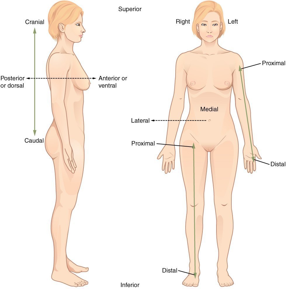
Anatomical position is the fixed reference: upright, face forward, upper limbs at the sides, palms forward. It applies even when a person is lying down, and right and left belong to the person described, not the viewer.
Anatomical directional terms on a standing body show superior toward the head, inferior toward the feet, medial toward the midline, and lateral away from it. OpenStax College. Anatomy & Physiology, Connexions Web site. http://cnx.org/content/col11496/1.6/, Jun 19, 2013.. Directional Terms.jpg. CC BY 3.0. Displayed at reduced size; anatomical labels unchanged.Negative feedback during environmental heating. A rise in internal temperature is detected, compared with the regulated range, and opposed by effectors that increase heat loss. Fever differs: inflammatory signals raise the defended temperature itself. Original diagram, McNova Enterprises, 2026; free to reuse in this course.Text description of this figure
Four boxes form a loop. Box 1, stimulus: internal temperature rises above its range. An afferent signal goes to box 2, sensors: thermoreceptors in skin and hypothalamus. Their information is compared in box 3, integration: the hypothalamus compares input with the regulated range. An efferent signal goes to box 4, effectors: sweat glands secrete and skin blood vessels dilate. The response, heat loss by evaporation and radiation from skin, points back to the stimulus box. A central label states that negative feedback means the response opposes the original change. A note adds that humid air limits evaporation.
Lesson 1.1
Anatomical language and body organization
Distinguish anatomy physiology and pathophysiology
Describe six levels of organization
Apply anatomical position and directional terms
Anatomy concerns the form and relationships of body structures. Physiology concerns their functions. Pathophysiology examines altered function associated with disease or injury. These questions connect: a thin exchange surface supports diffusion, while thickening that surface may reduce exchange. Naming a structure and explaining a mechanism are separate tasks.
Chemical components form cells; related cells and extracellular material form tissues; tissues form organs; organs cooperate in systems; systems sustain the organism. A kidney is an organ containing several tissues, rather than a single tissue type. Organ systems are useful ways to organize study, but their functions overlap. Temperature regulation requires nervous signals, skin blood vessels, sweat glands, and metabolic heat production.
Use anatomical position as the reference even when a person is lying down: upright, face forward, upper limbs at the sides, palms forward. Right and left belong to the person described. Medial means toward the midline and lateral means away. Proximal and distal describe distance from a limb attachment or another stated origin. Superficial and deep compare distance from the surface. The elbow is proximal to the wrist; the sternum is medial to the shoulders. A relationship needs two structures and a reference position.
Apply the idea
Write six directional comparisons involving the trunk and limbs. Classify cell, skin, epithelial tissue, and nervous system by organizational level.
Compare your reasoning
Skin is an organ, epithelial tissue a tissue, a cell a cellular unit, and the nervous system an organ system. Accept directional statements that name both structures and use anatomical position.
Tissues form organs, and organs cooperate in systems. A kidney contains several tissue types, so it is an organ rather than a single tissue. The urinary system is the organ system it belongs to.
Lesson 1.2
Planes cavities and imaging orientation
Compare sagittal frontal and transverse sections
Locate major body cavities
Explain serous membrane relationships
A sagittal section divides left and right portions; only a midsagittal section divides equal halves. A frontal section divides anterior and posterior portions. A transverse section divides superior and inferior portions. A section may be oblique to these reference planes. When interpreting an unfamiliar image, find its orientation markers before naming a side.
The dorsal cavity includes the cranial and vertebral cavities surrounding the central nervous system. The ventral cavity includes thoracic and abdominopelvic divisions, separated by the diaphragm. Each lung occupies a pleural compartment. The heart lies within the pericardial sac in the mediastinum. The abdominal and pelvic regions are continuous; a drawn boundary between them is not a physical wall.
Serous membranes have a parietal layer associated with the cavity wall and a visceral layer associated with the organ. A thin fluid film reduces friction between surfaces. Do not confuse the pleural cavity, a potential space between membrane layers, with the whole space occupied by the lung. In axial clinical images, display conventions can place the patient's right on the viewer's left. The image's labels take priority over an assumption about screen position.
Apply the idea
Describe the route from skin to the surface of a lung using superficial, deep, parietal, and visceral. Explain which section would show both knees in superior and inferior portions.
Compare your reasoning
A transverse section divides superior and inferior portions. The parietal pleura lines the thoracic wall and visceral pleura covers the lung; the intervening pleural space contains a small fluid film.
Display conventions for axial images can place the patient's right on the viewer's left. The image's labels take priority over assumptions about screen position.
Lesson 1.3
Homeostatic control and feedback
Trace stimulus sensor integration and effector
Contrast negative and positive feedback
Distinguish fever from environmental heating
Homeostasis maintains internal conditions within ranges compatible with cell function. Variables fluctuate: regulation is ongoing rather than a fixed reading. A negative feedback response opposes a disturbance. A sensor detects a variable, an integrating process compares information with the regulated range, and an effector changes activity. Feedback describes the direction of the response, not whether the response is pleasant or harmful.
When internal temperature rises during environmental heating, hypothalamic pathways can increase sweating and skin blood flow. Evaporation removes heat when conditions permit. High humidity limits evaporation, and sweating without evaporation cools less effectively. Fever differs because inflammatory signals raise the defended temperature. Chills may occur while the body is below that new target. This distinction explains why similar temperature readings can arise through different mechanisms.
Positive feedback reinforces a process until an endpoint interrupts it. Cervical stretch and oxytocin release during childbirth illustrate the pattern. Positive feedback is not the usual mechanism for keeping a variable near a target. In disease, a deviation can reflect a changed input, a sensing problem, a signaling failure, or limited effector function. Observing a deviation alone does not identify which part failed.
Apply the idea
Draw separate negative feedback loops for heat exposure and a thermostat. Name one limit of the thermostat analogy. Explain why sweating is not proof that body temperature is safe.
Compare your reasoning
Both models include sensing, integration, and an opposing response. Human targets and responses vary with context and interact with other systems. Sweating may fail to remove enough heat in humid conditions.
In fever, the regulated (defended) temperature is raised. Chills and shivering can occur while the body is below that new target. This is why similar temperature readings can arise through different mechanisms.
Watch for these errors
Common error: Negative feedback means the response is harmful or unpleasant.
Better reasoning: Negative describes direction only: the response opposes the change. Sweating that lowers a rising temperature is negative feedback even though it feels uncomfortable.
Common error: Left and right on a diagram or scan are the viewer's left and right.
Better reasoning: Right and left always belong to the person described. Check orientation labels on any image before naming a side.
Draw to learn: a negative feedback loop
Producing your own labeled sketch from memory, then checking it, is a generative activity that supports later recall. Paper, a tablet, or McNova Drawing all work.
Without looking at the lesson, draw a loop for body temperature during environmental heating. Label the stimulus, sensor, integrating center, effector, and response, and draw the arrow that shows the response opposing the change.
Self-check after drawing
Virtual lab
Temperature feedback model
Investigate Does stronger negative feedback reduce a simulated temperature deviation?
Synthetic teaching data · Estimated work is included in this module laboratory allocation · 15 point report
Procedure
Use the supplied model data; no exercise or body temperature measurement is required.
Identify elapsed time as the independent variable, temperature as the dependent variable, and the same initial heat load as a controlled condition.
Plot both response series on one graph with units.
Calculate change from the initial value and compare the final distance from the model target of 37.0 °C.
Repeat the comparison using the interactive feedback model and record its assumptions.
Time min
Feedback active °C
Feedback reduced °C
0
38.2
38.2
2
37.8
38.1
4
37.5
38.0
6
37.3
37.9
8
37.2
37.8
Analysis and submission
Which series returns closer to target?
Calculate the reduction in deviation in each series.
Name a measurement uncertainty and a model limitation.
Submit an evidence sheet with a prediction, the provided observations, your graph or text equivalent, calculations with units, and a limitation. In public practice, save it on your own device.
Model limits The model omits humidity, metabolic heat, skin perfusion, and individual variation. It does not establish safe temperatures or a treatment response.
Expected interpretation and calculation check
Active feedback reduces deviation from 1.2 to 0.2 °C, a 1.0 °C reduction. Reduced feedback reduces it from 1.2 to 0.8 °C, a 0.4 °C reduction. These are invented model readings, not patient observations.
Feedback model
Model equation: next deviation = current deviation × (1 − response fraction). Five equal steps; initial temperature 38.2 °C; target 37.0 °C; no new heat input. This is a mathematical model, not a prediction for a person.
Clinical reasoning
A rising temperature
A fictional model is heated while its sweating pathway remains active. Its temperature continues to rise.
Your task Explain two reasons why an active effector may fail to restore a variable. Separate observation from proposed mechanism.
Use structure → normal function → disruption → observable consequence → uncertainty. Suggested response 150 to 250 words; cases remain fictional.
Case discussion
The observation is a rising temperature despite an active pathway. Heat input may exceed heat loss, or evaporation may be limited. The case does not establish fever or a particular illness.
Explain it in your own words
Two people both have a temperature of 38.5 °C. One has a fever and one is working in a hot, humid room. Explain why their bodies respond differently.
Compare with a model explanation
In the hot room, temperature rose because heat gain exceeded heat loss; the defended range has not changed, so the hypothalamus drives sweating and skin vasodilation to lose heat. High humidity limits evaporation, so cooling may lag. In fever, inflammatory signals raise the defended temperature itself. Until the body reaches that new target, it acts as if it is too cold, which can include shivering and skin vasoconstriction. The same reading reflects two different mechanisms.
Close the module
1. Free recall (3 minutes, book closed). List every directional term, plane, and body cavity you can remember, then write the five parts of a feedback loop in order.
2. Rate your confidence on each objective (1 not yet, 4 confident), then take the checkpoint and compare. Large gaps between confidence and score show where to restudy.
Objective
1
2
3
4
Anatomical language and body organization
Planes cavities and imaging orientation
Homeostatic control and feedback
3. Muddiest point. Write one sentence naming what is still least clear, and plan when you will revisit it (for example, in two days and again in a week).
Module checkpoint
Four questions worth 5 practice points each. Use the course player for saved attempts, or use the immediate practice feedback below.
Discussion or reflection
Choose one observation from this module. Explain it in 120 to 180 words using a mechanism, compare a plausible alternative, and state what evidence would distinguish them. In a group, respond to one peer using a source and a question. In public self-study, write the alternative yourself. No external account or public posting is required.
Connect bonds, water, and pH to how cells work, and explain how enzymes and ATP let cells do chemical work.
Suggested sequence (about 150 minutes of focused study, plus practice)
Make a prediction (2 minutes)
Lesson: Atoms bonds and water, then its retrieval check
Lesson: Acids bases buffers and pH, then its retrieval check
Lesson: Biomolecules enzymes and ATP, then its retrieval check
Watch for common errors and complete the drawing task
Virtual lab: Buffer comparison
Clinical reasoning case: Reduced oxygen delivery
Explain it in your own words
Spaced review and close the module
Module checkpoint (four questions)
By the end you should be able to
Distinguish ionic covalent and hydrogen interactions
Connect water polarity to biological behavior
Explain electrolytes in solution
Interpret the logarithmic pH scale
Explain how a buffer limits pH change
Distinguish a model solution from a clinical specimen
Match four biomolecule groups to functions
Explain enzyme specificity and regulation
Link ATP regeneration to cellular work
Before you start: make a prediction
Guessing is expected here. Attempting a question before the lesson tends to make the explanation that follows easier to remember, even when the first answer is wrong. This item is not scored.
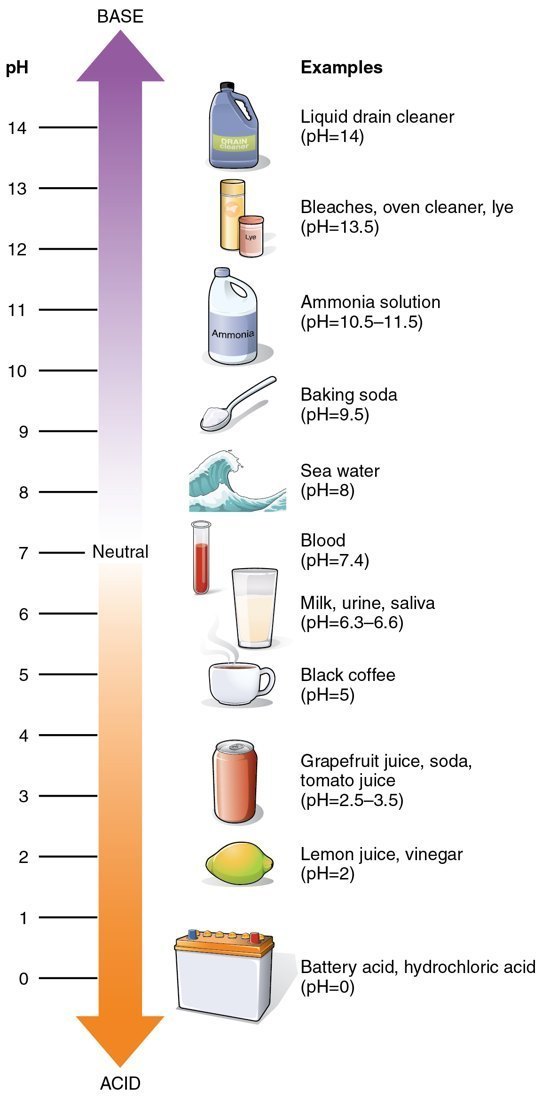
The pH scale is logarithmic. Each one-unit drop is a tenfold rise in hydrogen ion concentration, so a two-unit drop is 10 × 10 = 100 times more.
The logarithmic pH scale compares acidic, neutral, and alkaline solutions; lower pH corresponds to greater hydrogen ion concentration. OpenStax College. 216 pH Scale-01.jpg. CC BY 3.0. Displayed at reduced size; anatomical labels unchanged.The pH scale is logarithmic. Each pH unit is a tenfold change in hydrogen ion concentration, so a two-unit drop is a hundredfold rise. The narrow band marks the commonly taught arterial blood interval. Original diagram, McNova Enterprises, 2026; free to reuse in this course.Text description of this figure
A horizontal pH scale runs from 0 at the left, labeled more acidic, to 14 at the right, labeled more basic. Beneath selected pH values the hydrogen ion concentration is shown as powers of ten, from 10 to the 0 at pH 0 to 10 to the minus 14 at pH 14. A narrow band at 7.35 to 7.45 marks arterial blood. A curved arrow from pH 7 to pH 5 is labeled 10 times 10 equals 100 times more hydrogen ions. A footnote states that each one-unit drop in pH means ten times more hydrogen ions and that pH 7 is neutral at 25 degrees Celsius.
Lesson 2.1
Atoms bonds and water
Distinguish ionic covalent and hydrogen interactions
Connect water polarity to biological behavior
Explain electrolytes in solution
An atom contains a nucleus with protons and neutrons, surrounded by electrons. Proton number identifies the element. Isotopes have different neutron numbers. Ions form when atoms or groups gain or lose electrons, producing net charge. In a solution, dissolved ions can carry electrical current; these solutes are called electrolytes.
Covalent bonds involve shared electrons. Unequal sharing produces polar covalent bonds. Ionic attraction occurs between oppositely charged ions, although water can separate many ions into solution. Hydrogen bonds are weaker attractions between suitable polar groups; they help stabilize protein and nucleic acid arrangements and interactions among water molecules. Breaking a hydrogen bond is not the same event as splitting a water molecule.
Water's polarity helps dissolve many charged and polar substances. Its heat capacity limits rapid temperature changes, and evaporation transfers heat away from a surface. Nonpolar molecules interact less favorably with water. This behavior contributes to membrane organization. Describe the specific interaction rather than saying water dissolves everything: membrane lipids form barriers in part because their nonpolar regions avoid contact with water.
Apply the idea
Compare sodium chloride dissolved in water with a membrane phospholipid. State which parts interact with water and why.
Compare your reasoning
Water stabilizes separated sodium and chloride ions through charge interactions. A phospholipid has a polar head that interacts with water and nonpolar tails that associate away from water.
Nonpolar molecules interact less favorably with water. Membrane lipids form barriers in part because their nonpolar regions avoid contact with water, which organizes the bilayer.
Lesson 2.2
Acids bases buffers and pH
Interpret the logarithmic pH scale
Explain how a buffer limits pH change
Distinguish a model solution from a clinical specimen
An acid can donate hydrogen ions; a base can accept them. For dilute aqueous solutions, pH expresses the negative base-ten logarithm of hydrogen ion concentration. A decrease of one pH unit corresponds to a tenfold increase in hydrogen ion concentration. A change from pH 7 to pH 5 is a hundredfold increase, not a twofold increase.
A buffer contains a weak acid and its conjugate base, or the corresponding base system. It can accept or release hydrogen ions and limit pH change when small quantities of acid or base are added. Buffer capacity is finite. A buffer does not prevent every change, and its action does not remove the need for regulation by the lungs and kidneys.
Arterial blood pH is commonly described as approximately 7.35 to 7.45. Reference intervals depend on specimen and laboratory context; this interval is not interchangeable with urine or stomach pH. Changes in hydrogen ion concentration can alter protein charge, enzyme activity, and electrical behavior. The chemical mechanism belongs in this course; clinical interpretation also requires carbon dioxide, bicarbonate, sampling conditions, and the person's circumstances.
Apply the idea
Calculate the hydrogen ion concentration ratio for pH 6 versus pH 8. Explain why a buffer can eventually fail to hold pH nearly constant.
Compare your reasoning
The pH 6 solution has 100 times the hydrogen ion concentration of the pH 8 solution. Buffer components are used as acid or base is added, so protection is limited by their available amounts.
A buffer accepts or releases hydrogen ions to limit pH change, but it does not prevent every change. In the course buffer lab, the buffer pH still changed by 0.7 units.
Lesson 2.3
Biomolecules enzymes and ATP
Match four biomolecule groups to functions
Explain enzyme specificity and regulation
Link ATP regeneration to cellular work
Carbohydrates include sugars and their polymers; they participate in energy use, storage, and cell recognition. Lipids include triglycerides, phospholipids, and steroids. Their functions include fuel storage, membranes, and signaling. Proteins consist of amino acid chains whose folding supports roles such as transport, catalysis, contraction, and structural support. Nucleic acids contain nucleotides: DNA stores hereditary information, and several forms of RNA participate in its expression.
Enzymes lower activation energy and increase reaction rates without being consumed by each reaction cycle. They do not change the overall free energy difference between reactants and products. Substrate binding depends on molecular arrangement and chemical conditions. Temperature and pH can alter enzyme behavior; heating does not always increase activity because protein structure can be disrupted.
ATP transfers usable chemical energy through coupled reactions. Cells regenerate ATP continually. Glycolysis occurs in the cytosol; mitochondrial processes account for much ATP production under aerobic conditions. Oxygen serves as the final electron acceptor in the electron transport chain. ATP yield varies with conditions, so one universal exact yield is misleading. Reduced oxygen supply can impair oxidative ATP production, affecting pumps, cell volume, and contraction. ATP also participates in muscle relaxation, not only force production.
Apply the idea
Build a chain from reduced oxidative ATP production to altered ion pumping and cell swelling. Identify a step that also depends on membrane permeability.
Compare your reasoning
Reduced ATP can impair sodium potassium pumping; ionic gradients may dissipate and effective intracellular solute distribution can favor water entry. The magnitude depends on ion pathways and other transport processes.
Enzymes increase reaction rates by lowering activation energy. They do not change the overall free energy difference, and heating can disrupt protein structure and reduce activity.
Watch for these errors
Common error: pH 5 is only a little more acidic than pH 7, about twice as acidic.
Better reasoning: The scale is logarithmic. pH 5 has 100 times the hydrogen ion concentration of pH 7.
Common error: ATP is needed for muscle contraction but not for relaxation.
Better reasoning: ATP is also needed for myosin to detach from actin and for pumps that remove calcium, so a shortage can impair relaxation.
Draw to learn: the pH scale and a buffer
Producing your own labeled sketch from memory, then checking it, is a generative activity that supports later recall. Paper, a tablet, or McNova Drawing all work.
Draw a pH scale from 0 to 14. Mark neutral, the arterial blood interval, and show with arrows how many times more hydrogen ions there are at pH 6 and pH 5 than at pH 7. Beside it, sketch a buffer pair taking up an added hydrogen ion.
Self-check after drawing
Virtual lab
Buffer comparison
Investigate Which solution resists an acid challenge more effectively?
Synthetic teaching data · Estimated work is included in this module laboratory allocation · 15 point report
Procedure
Use only supplied simulated readings; do not mix household chemicals.
Keep initial sample volume and acid aliquot size constant in the model.
Plot pH against number of equal acid aliquots.
Calculate total pH change for each solution.
Explain why a pH difference is not a linear measure of hydrogen ion concentration.
Acid aliquots
Water model pH
Buffer model pH
0
7.0
7.0
1
5.0
6.9
2
4.7
6.8
3
4.5
6.6
4
4.4
6.3
Analysis and submission
Which condition has greater resistance?
How much does each pH decrease?
Why should you avoid calculating a percent acid concentration from these pH numbers?
Submit an evidence sheet with a prediction, the provided observations, your graph or text equivalent, calculations with units, and a limitation. In public practice, save it on your own device.
Model limits The data are synthetic; acid strength, buffer species, and molar concentrations are not specified. Do not infer clinical buffering capacity.
Expected interpretation and calculation check
Water decreases 2.6 pH units and the buffer 0.7. The buffer better resists the challenge, but its pH still changes. A logarithmic scale cannot be treated as a linear concentration scale.
Clinical reasoning
Reduced oxygen delivery
A hypothetical cell receives less oxygen but still has glucose. ATP-dependent transport declines.
Your task Explain why glucose availability alone does not ensure the usual aerobic ATP supply.
Use structure → normal function → disruption → observable consequence → uncertainty. Suggested response 150 to 250 words; cases remain fictional.
Case discussion
Glycolysis can continue under some conditions, but electron transport requires oxygen. Lower oxidative ATP production may limit energy-dependent transport. The case does not identify a cause of reduced oxygen delivery.
Explain it in your own words
Explain how a fall in oxygen supply could cause a cell to swell. Name each step between oxygen and cell volume.
Compare with a model explanation
Oxygen is the final electron acceptor in the electron transport chain, so less oxygen reduces oxidative ATP production. The sodium potassium pump uses ATP to move three sodium ions out and two potassium ions in. With less ATP, the pump slows and sodium builds up inside the cell. More solute inside draws water in by osmosis, so the cell swells. Each step is a link that could be tested separately.
Spaced review
These items return to earlier modules on purpose. Retrieving older material after a delay, mixed with new topics, strengthens long-term retention and helps you choose the right method when problem types are mixed.
A frontal plane separates anterior and posterior portions; a transverse plane separates superior and inferior portions.
Close the module
1. Free recall (3 minutes, book closed). Write the four biomolecule groups with one function each, then explain in two sentences what a buffer does and does not do.
2. Rate your confidence on each objective (1 not yet, 4 confident), then take the checkpoint and compare. Large gaps between confidence and score show where to restudy.
Objective
1
2
3
4
Atoms bonds and water
Acids bases buffers and pH
Biomolecules enzymes and ATP
3. Muddiest point. Write one sentence naming what is still least clear, and plan when you will revisit it (for example, in two days and again in a week).
Module checkpoint
Four questions worth 5 practice points each. Use the course player for saved attempts, or use the immediate practice feedback below.
Discussion or reflection
Choose one observation from this module. Explain it in 120 to 180 words using a mechanism, compare a plausible alternative, and state what evidence would distinguish them. In a group, respond to one peer using a source and a question. In public self-study, write the alternative yourself. No external account or public posting is required.
Explain how cells make and export proteins, control what crosses the membrane, and divide under checkpoint control.
Suggested sequence (about 150 minutes of focused study, plus practice)
Make a prediction (2 minutes)
Lesson: Organelles and protein production, then its retrieval check
Lesson: Membrane transport and tonicity, then its retrieval check
Lesson: Signals gene expression and cell division, then its retrieval check
Watch for common errors and complete the drawing task
Virtual lab: Osmosis and cell volume
Clinical reasoning case: A cell becomes swollen
Explain it in your own words
Spaced review and close the module
Module checkpoint (four questions)
By the end you should be able to
Identify major organelle functions
Trace a secreted protein through the cell
Connect structure to intracellular transport
Compare passive and active transport
Predict water movement with nonpenetrating solutes
Distinguish osmolarity from tonicity
Distinguish receptor signaling from transport
Order the cell cycle
Explain why uncontrolled growth needs more than a mitosis label
Before you start: make a prediction
Guessing is expected here. Attempting a question before the lesson tends to make the explanation that follows easier to remember, even when the first answer is wrong. This item is not scored.
Pure water has lower solute concentration than the cell, so it is hypotonic. Net water movement is inward and the cell swells. Module 3 explains why tonicity depends on solutes that cannot cross the membrane.
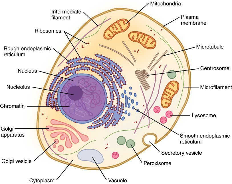
A labeled animal cell locates nucleus, endoplasmic reticulum, Golgi apparatus, mitochondria, and membrane-bound compartments within the plasma membrane. OpenStax. 0312 Animal Cell and Components.jpg. CC BY 4.0. Displayed at reduced size; anatomical labels unchanged.Tonicity and animal cell volume. Tonicity describes the effect of a solution on cell volume and depends on solutes that do not rapidly cross the membrane. The arrows show the initial direction of net water movement in a simplified model. Original diagram, McNova Enterprises, 2026; free to reuse in this course.Text description of this figure
Three panels each show a cell compared with a dashed circle marking its starting size. Left panel, hypotonic outside: lower nonpenetrating solute outside than inside; arrows point inward and the cell is larger, labeled net water moves in, cell swells. Middle panel, isotonic outside: equal effective solute; paired arrows point both ways and the cell matches its starting size, labeled no net volume change, water still moves both ways. Right panel, hypertonic outside: higher nonpenetrating solute outside; arrows point outward and the cell is smaller, labeled net water moves out, cell shrinks.
Lesson 3.1
Organelles and protein production
Identify major organelle functions
Trace a secreted protein through the cell
Connect structure to intracellular transport
The plasma membrane encloses the cell, and cytoplasm includes cytosol and organelles. The nucleus contains most cellular DNA. Ribosomes translate messenger RNA into polypeptides. Free ribosomes and ribosomes associated with rough endoplasmic reticulum perform the same translation chemistry, but the location helps direct the destination of the resulting protein.
A secreted protein typically enters the rough endoplasmic reticulum during synthesis, passes through processing and sorting steps that include the Golgi apparatus, and travels in vesicles to the membrane. Exocytosis releases vesicle contents outside the cell. Lysosomes contain enzymes that break down selected materials; peroxisomes participate in oxidative reactions. Mitochondria support aerobic energy metabolism and contain their own small genome.
The cytoskeleton includes microfilaments, intermediate filaments, and microtubules. It supports cell shape, movement, division, and directed intracellular transport. Organelles cooperate rather than acting as isolated compartments. A defect in protein sorting can change secretion even when transcription and translation continue. A mature human erythrocyte lacks a nucleus and mitochondria, reminding us that a general cell diagram is a model rather than a description of every cell.
Apply the idea
Trace a glandular protein from DNA information to release. Identify two different cellular defects that could reduce secretion.
Compare your reasoning
DNA transcription produces RNA; ribosomes translate protein; rough ER, Golgi, and vesicles process and direct it to exocytosis. Reduced transcription, defective folding, sorting, or vesicle transport could each reduce release.
A secreted protein typically enters the rough ER during synthesis, passes through processing and sorting steps that include the Golgi apparatus, and leaves the cell by exocytosis.
Lesson 3.2
Membrane transport and tonicity
Compare passive and active transport
Predict water movement with nonpenetrating solutes
Distinguish osmolarity from tonicity
The lipid bilayer is selectively permeable. Small nonpolar molecules can cross by simple diffusion. Ions and many polar substances need channels or carriers. Facilitated diffusion moves a substance down its electrochemical gradient without directly supplying metabolic energy to that movement. Active transport moves substances against a gradient through energy coupling. The sodium potassium ATPase transports three sodium ions outward and two potassium ions inward per ATP cycle.
Osmosis is net water movement across a selectively permeable boundary. In a simplified model with nonpenetrating solute, water moves toward the compartment with the greater effective solute concentration. Osmolarity counts dissolved particles per liter. Tonicity describes a solution's effect on cell volume and depends on solutes that do not rapidly cross the membrane. Equal osmolarity does not guarantee equal tonicity if solutes have different permeability.
An animal cell placed in a hypotonic solution tends to gain water, while one in a hypertonic solution tends to lose water. In an isotonic solution, no sustained net volume change is expected from that comparison. Water molecules still move both ways. Filtration differs: a hydrostatic pressure difference contributes to movement of fluid and solutes through a barrier. These models explain physical processes; they do not prescribe intravenous fluids.
Apply the idea
A water-permeable membrane separates 300 mOsm/L nonpenetrating solute on the left from 200 on the right. Predict initial water movement. State what changes if the solute also crosses rapidly.
Compare your reasoning
Initial net water movement is toward the left. Rapid solute entry can change the effective osmotic difference over time, so the original sustained-volume prediction may fail.
Osmolarity counts dissolved particles. Tonicity describes the effect on cell volume and depends on solutes that do not rapidly cross the membrane. A penetrating solute can enter the cell, so equal osmolarity can still change volume.
Lesson 3.3
Signals gene expression and cell division
Distinguish receptor signaling from transport
Order the cell cycle
Explain why uncontrolled growth needs more than a mitosis label
A cell responds to a signal only if it has the relevant receptor and downstream machinery. A membrane receptor can bind a signal without carrying it across the bilayer. Some lipid-soluble signals act through intracellular receptors. Signal transduction changes events within the cell, such as enzyme activity or gene expression. Receptor presence, concentration, and cellular context affect the response.
DNA replication occurs during S phase of interphase. G1 and G2 provide periods of growth and preparation; cells may also enter a nondividing state often called G0. Mitosis separates duplicated chromosomes into daughter nuclei, and cytokinesis divides cytoplasm. Prophase, metaphase, anaphase, and telophase describe chromosome behavior, not DNA synthesis. A checkpoint can delay progression when conditions are unsuitable or DNA is damaged.
Apoptosis is regulated cell death that supports tissue maintenance and development. Aging involves changes across cells and tissues; it is not equivalent to every cell simply reaching the same division count. Cancer involves disrupted controls of growth and survival, often with accumulated genetic changes. A microscopic mitotic figure shows a dividing cell; it alone does not establish malignancy, since normal renewing tissues also divide.
Apply the idea
Put DNA replication, chromosome alignment, chromosome separation, and cytokinesis in order. Explain why many dividing cells can occur during normal repair.
Compare your reasoning
Replication precedes mitosis; chromosomes align in metaphase, sister chromatids separate in anaphase, and cytokinesis divides the cytoplasm. Normal repair includes regulated cell proliferation.
Normal renewing tissues also divide, so a mitotic figure alone does not establish malignancy. DNA replication happens in S phase of interphase, not during mitosis.
Watch for these errors
Common error: In an isotonic solution water stops moving across the membrane.
Better reasoning: Water molecules still move both ways. Isotonic means there is no net volume change, not that movement stops.
Common error: DNA is copied during mitosis.
Better reasoning: DNA replication occurs in S phase of interphase. Mitosis separates chromosomes that were already duplicated.
Draw to learn: the secretory pathway
Producing your own labeled sketch from memory, then checking it, is a generative activity that supports later recall. Paper, a tablet, or McNova Drawing all work.
Draw a cell with nucleus, rough ER, Golgi apparatus, and plasma membrane. Use numbered arrows to trace a secreted protein from its mRNA to its release outside the cell.
Self-check after drawing
Virtual lab
Osmosis and cell volume
Investigate How does external nonpenetrating solute concentration affect initial water movement?
Synthetic teaching data · Estimated work is included in this module laboratory allocation · 15 point report
Procedure
Assume an initial intracellular concentration of 300 mOsm/L and equal initial cell volumes.
Treat all listed solutes as nonpenetrating during the observation period.
Predict each response before examining the provided relative volumes.
Calculate percent volume change as 100 × (final minus initial) divided by initial.
Use the osmosis model to vary external concentration and explain the difference between directional prediction and measured volume.
External mOsm/L
Initial relative volume
Final relative volume
200
1.0
1.25
300
1.0
1.0
400
1.0
0.82
Analysis and submission
Classify all three conditions.
Calculate each volume change.
Why does this dataset not prove an exact inverse concentration-volume law for living cells?
Submit an evidence sheet with a prediction, the provided observations, your graph or text equivalent, calculations with units, and a limitation. In public practice, save it on your own device.
Model limits Synthetic final volumes are illustrative, not derived from a calibrated cell model. Real cells may regulate volume and solutes may cross. No tissue or human sampling is needed.
Expected interpretation and calculation check
The conditions are hypotonic, isotonic, and hypertonic relative to the cell. Changes are +25%, 0%, and −18%. Water moves initially inward, equally in both directions with no net volume change, and outward, respectively.
Osmosis direction model
Internal concentration is initially 300 mOsm/L. Water can cross; solute cannot. The model predicts initial direction only, not final cell size or a fluid prescription.
Clinical reasoning
A cell becomes swollen
A fictional cell has impaired ATP regeneration and gradually loses control of its ion distribution. Water enters and cell volume rises.
Your task Connect energy supply, membrane transport, solute distribution, and water movement without claiming that all swelling has this cause.
Use structure → normal function → disruption → observable consequence → uncertainty. Suggested response 150 to 250 words; cases remain fictional.
Case discussion
ATP-dependent transport can fail to maintain ion gradients. A changed distribution of effectively nonpenetrating particles can favor water entry. Swelling alone does not identify the initial defect.
Explain it in your own words
Explain why a cell placed in a solution of urea can swell even when the urea solution has the same osmolarity as the cell.
Compare with a model explanation
Osmolarity counts all particles, but tonicity depends only on solutes that stay outside. Urea crosses the membrane, so it moves into the cell over time. As urea enters, the effective solute inside rises compared with outside, and water follows by osmosis. The cell gains water and swells. The solution started iso-osmotic but behaves as hypotonic, which is why osmolarity and tonicity must be kept separate.
Spaced review
These items return to earlier modules on purpose. Retrieving older material after a delay, mixed with new topics, strengthens long-term retention and helps you choose the right method when problem types are mixed.
Oxygen supports electron transport; reduced oxidative ATP supply can limit ATP-dependent transport.
Markers establish orientation. Location on a screen alone is insufficient to assign a patient side.
Close the module
1. Free recall (3 minutes, book closed). Name the organelles a secreted protein passes through, list the three tonicity conditions with the direction of net water movement, and order the cell cycle phases.
2. Rate your confidence on each objective (1 not yet, 4 confident), then take the checkpoint and compare. Large gaps between confidence and score show where to restudy.
Objective
1
2
3
4
Organelles and protein production
Membrane transport and tonicity
Signals gene expression and cell division
3. Muddiest point. Write one sentence naming what is still least clear, and plan when you will revisit it (for example, in two days and again in a week).
Module checkpoint
Four questions worth 5 practice points each. Use the course player for saved attempts, or use the immediate practice feedback below.
Discussion or reflection
Choose one observation from this module. Explain it in 120 to 180 words using a mechanism, compare a plausible alternative, and state what evidence would distinguish them. In a group, respond to one peer using a source and a question. In public self-study, write the alternative yourself. No external account or public posting is required.
Read tissue features as evidence for function, and explain why some injuries regenerate while others form scar.
Suggested sequence (about 150 minutes of focused study, plus practice)
Make a prediction (2 minutes)
Lesson: Epithelium and glands, then its retrieval check
Lesson: Connective muscle and nervous tissues, then its retrieval check
Lesson: Inflammation regeneration and fibrosis, then its retrieval check
Watch for common errors and complete the drawing task
Virtual lab: Digital tissue identification
Clinical reasoning case: Repair with reduced function
Explain it in your own words
Spaced review and close the module
Module checkpoint (four questions)
By the end you should be able to
Classify epithelium by layers and shape
Relate polarity and avascularity to function
Distinguish exocrine from endocrine secretion
Classify connective tissue by cells and matrix
Compare three muscle tissue types
Distinguish neurons from supporting glia
Explain the purpose and limits of inflammation
Distinguish regeneration from fibrosis
Relate blood supply and tissue properties to healing
Before you start: make a prediction
Guessing is expected here. Attempting a question before the lesson tends to make the explanation that follows easier to remember, even when the first answer is wrong. This item is not scored.
Fibrosis deposits connective tissue that restores continuity but may not reproduce glands, follicles, nerves, or the original architecture. Lesson 4.3 contrasts this with regeneration.
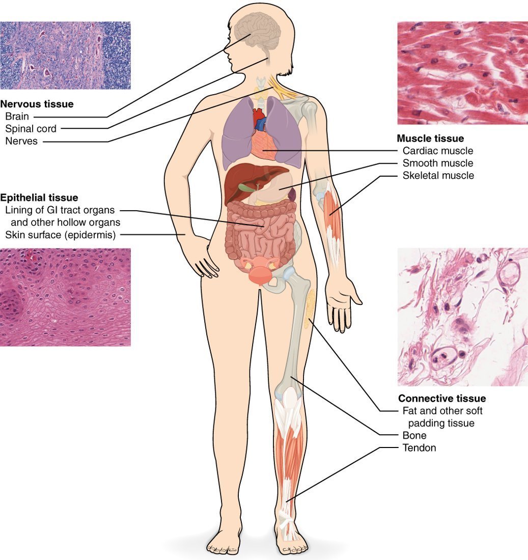
Four tissue families are illustrated by epithelial coverings, connective support, contractile muscle, and signaling nervous tissue. OpenStax College. 401 Types of Tissue.jpg. CC BY 3.0. Displayed at reduced size; anatomical labels unchanged.Two outcomes of tissue repair. Repair moves through overlapping events. Whether lost tissue regenerates or is replaced by scar depends on cell proliferative capacity, blood supply, scaffolding, extent of damage, and mechanical conditions. Original diagram, McNova Enterprises, 2026; free to reuse in this course.Text description of this figure
A row of four boxes shows injury, then bleeding control with a clot forming a temporary seal, then inflammation with fluid, proteins, and immune cells entering, then tissue formation with new cells, vessels, and matrix. Two arrows branch from tissue formation. One leads to regeneration, where lost cells are replaced with the original tissue pattern, more likely with dividing cells, good blood supply, and intact scaffold. The other leads to fibrosis or scar, where collagen restores continuity but glands, nerves, and original architecture may not return. Both lead to remodeling, where matrix is reorganized over time. Notes state that many injuries heal by a mix of both routes, phases overlap, and persistent inflammation can damage tissue.
Lesson 4.1
Epithelium and glands
Classify epithelium by layers and shape
Relate polarity and avascularity to function
Distinguish exocrine from endocrine secretion
Epithelium covers surfaces, lines cavities, and forms glands. Cells are closely associated, with relatively little extracellular matrix. The apical surface faces a lumen or external surface, while the basal surface attaches to a basement membrane. Epithelium lacks its own blood vessels; nutrients diffuse from nearby connective tissue. Rapid renewal depends on the tissue and its stem cell population.
Name an epithelium by its number of layers and the shape of cells at the exposed surface. Simple squamous epithelium provides a thin surface suited to exchange. Stratified squamous epithelium resists abrasion. Cuboidal and columnar cells often support secretion or absorption. Pseudostratified epithelium appears multilayered because nuclei occupy different heights, but all cells contact the basement membrane. Transitional epithelium changes appearance as the urinary tract wall stretches.
Exocrine glands release products through ducts onto surfaces or into lumens. Endocrine glands release hormones into the interstitial environment, from which they enter circulation. A gland may include different cell populations and secretion routes. On a section, apparent layer number can be distorted by the angle of cutting. Use more than one field and consider where the free surface and basement membrane lie before classifying.
Apply the idea
Compare a thin exchange lining with a surface exposed to repeated abrasion. Predict the epithelial arrangements and give a reason for each.
Compare your reasoning
A thin simple squamous lining favors diffusion; multiple layers of stratified squamous cells protect against abrasion. Classification must still be verified from actual tissue features.
Pseudostratified epithelium appears multilayered because nuclei sit at different heights, but all cells contact the basement membrane, so it is a single layer.
Lesson 4.2
Connective muscle and nervous tissues
Classify connective tissue by cells and matrix
Compare three muscle tissue types
Distinguish neurons from supporting glia
Connective tissue is defined in part by its extracellular matrix, which includes ground substance and fibers. Loose connective tissue supports exchange and flexible packing. Dense regular connective tissue has aligned collagen suited to tension mainly in one direction; dense irregular tissue resists forces in several directions. Cartilage has a firm hydrated matrix and lacks its own blood vessels. Bone has mineralized matrix. Blood is connective tissue with a fluid matrix.
Skeletal muscle fibers are long, striated, and usually multinucleated, with nuclei near the periphery. Cardiac muscle cells are striated and branched, connected through intercalated discs. Smooth muscle cells lack visible striations in routine light microscopy and usually have one central nucleus. These visible features support identification, but section orientation can hide shape or striation.
Neurons receive and transmit signals; glial cells support many aspects of neural function, including myelination, chemical balance, and immune surveillance. Nervous tissue contains more than neurons. A tissue image should be described before it is named: note cell arrangement, matrix, nuclei, fibers, and any free surface. Record confidence and the feature that would resolve uncertainty rather than guessing from color alone.
Apply the idea
Create a four-row matrix for epithelial, connective, muscle, and nervous tissue. Include one visible feature, function, and common identification error per row.
Compare your reasoning
Accept tightly packed epithelium, matrix-rich connective tissue, contractile muscle morphology, and neuronal processes with glia. Color alone and a single uncertain feature are insufficient identifiers.
Cardiac muscle cells are striated and branched and connect through intercalated discs. Skeletal fibers are long and multinucleated; smooth muscle lacks visible striations.
Lesson 4.3
Inflammation regeneration and fibrosis
Explain the purpose and limits of inflammation
Distinguish regeneration from fibrosis
Relate blood supply and tissue properties to healing
Injury releases signals that recruit an inflammatory response. Changes in local blood flow and vessel permeability support the movement of fluid, proteins, and immune cells into the affected area. Redness, warmth, swelling, discomfort, and altered function can accompany inflammation. Their appearance varies with location, depth, and skin pigmentation. Inflammation can arise without infection, and infection cannot be established from redness alone.
Repair includes overlapping events rather than a rigid sequence with identical timing: control of bleeding, inflammation, tissue formation, and remodeling. Regeneration replaces lost cells with cells that restore the original tissue pattern when conditions permit. Fibrosis deposits connective tissue that may restore continuity while changing mechanical behavior and function. Some injuries heal through both processes.
Cell proliferative capacity, blood supply, extracellular scaffolding, extent of damage, and mechanical conditions affect the outcome. Cartilage generally repairs less readily than well vascularized tissues. A scar can close a defect without reproducing glands, nerve arrangements, or the original architecture. Persistent inflammation can damage tissue as well as accompany repair. A case explanation should identify the observed change, a plausible mechanism, and what evidence is still missing.
Apply the idea
Explain why closure of a skin defect does not prove full recovery of sensation or mechanical strength. Compare one regenerative process with one fibrotic process.
Compare your reasoning
New connective tissue may bridge a defect while original appendages and neural patterns remain altered. Restored continuity and restored original structure are different outcomes.
Proliferative capacity, blood supply, scaffolding, extent of damage, and mechanics affect outcome. Cartilage lacks its own blood vessels and generally repairs less readily than well-vascularized tissues.
Watch for these errors
Common error: Redness and swelling mean an infection is present.
Better reasoning: Inflammation can occur without infection, and its appearance varies with location, depth, and skin pigmentation. Redness alone cannot establish infection.
Common error: A closed wound means the tissue has been fully restored.
Better reasoning: A scar can close a defect without rebuilding glands, nerves, or architecture, so continuity is not the same as full function.
Draw to learn: four tissue types side by side
Producing your own labeled sketch from memory, then checking it, is a generative activity that supports later recall. Paper, a tablet, or McNova Drawing all work.
From memory, sketch one example of epithelium, connective tissue, muscle, and nervous tissue. Under each, write the one visible feature you would use to identify it.
Self-check after drawing
Virtual lab
Digital tissue identification
Investigate Which observed tissue features support a classification?
Synthetic teaching data · Estimated work is included in this module laboratory allocation · 15 point report
Procedure
Inspect the tissue illustration and descriptions below before revealing the interpretation.
For each specimen, record at least two observed features without using the tissue name.
Assign a tissue family and a likely function.
Write a plausible alternative and name a feature needed to reject it.
Submit a four-specimen identification sheet and a 150-word uncertainty statement.
Specimen
Observed features
Task
A
One thin layer of flattened cells bordering a space
Classify lining
B
Parallel collagen bundles and sparse elongated nuclei
Classify matrix
C
Long striated fibers with peripheral multiple nuclei
Classify muscle
D
Cell bodies with branching processes among small nuclei
Classify neural tissue
Analysis and submission
What is the best classification for each?
Which description supports tension mainly in one direction?
Why is an illustration insufficient evidence of a clinical diagnosis?
Submit an evidence sheet with a prediction, the provided observations, your graph or text equivalent, calculations with units, and a limitation. In public practice, save it on your own device.
Model limits The included overview is a labeled illustration, not an unknown micrograph practical. A faculty practical should add verified unlabeled slides with scale and orientation; illustration identification cannot replace microscopy competency.
Expected interpretation and calculation check
A: simple squamous epithelium; B: dense regular connective tissue; C: skeletal muscle; D: nervous tissue. Parallel collagen supports directional tensile loading. These are teaching descriptions, not actual diagnostic slides.
Clinical reasoning
Repair with reduced function
A fictional healed skin wound has a firm scar and reduced local sensation.
Your task Explain how closure can coexist with altered function. Identify information needed before predicting recovery.
Use structure → normal function → disruption → observable consequence → uncertainty. Suggested response 150 to 250 words; cases remain fictional.
Case discussion
Fibrous repair can restore continuity without recreating the original neural distribution. Injury depth, nerve involvement, elapsed time, and examination findings are needed to discuss recovery.
Explain it in your own words
A tendon heals after a tear but feels stiffer and weaker than before. Explain why, using the terms regeneration and fibrosis.
Compare with a model explanation
Dense regular tendon tissue has aligned collagen suited to pulling in one direction and a limited blood supply. After a tear, repair can rely partly on fibrosis: new collagen restores continuity, but its arrangement may not match the original parallel pattern. Less organized collagen transmits force less well and can change stiffness. Regeneration would rebuild the original pattern, but limited blood supply and the extent of damage favor scar. Mechanical loading during healing also affects how collagen is remodeled.
Spaced review
These items return to earlier modules on purpose. Retrieving older material after a delay, mixed with new topics, strengthens long-term retention and helps you choose the right method when problem types are mixed.
DNA replication precedes mitosis and occurs during S phase of interphase.
A frontal plane separates anterior and posterior portions; a transverse plane separates superior and inferior portions.
Close the module
1. Free recall (3 minutes, book closed). List the four tissue families with one feature and one function each, then write the overlapping stages of repair in order.
2. Rate your confidence on each objective (1 not yet, 4 confident), then take the checkpoint and compare. Large gaps between confidence and score show where to restudy.
Objective
1
2
3
4
Epithelium and glands
Connective muscle and nervous tissues
Inflammation regeneration and fibrosis
3. Muddiest point. Write one sentence naming what is still least clear, and plan when you will revisit it (for example, in two days and again in a week).
Module checkpoint
Four questions worth 5 practice points each. Use the course player for saved attempts, or use the immediate practice feedback below.
Discussion or reflection
Choose one observation from this module. Explain it in 120 to 180 words using a mechanism, compare a plausible alternative, and state what evidence would distinguish them. In a group, respond to one peer using a source and a question. In public self-study, write the alternative yourself. No external account or public posting is required.
Link the layers and appendages of skin to barrier protection, heat exchange, vitamin D synthesis, and patterns of injury.
Suggested sequence (about 150 minutes of focused study, plus practice)
Make a prediction (2 minutes)
Lesson: Skin layers cells and appendages, then its retrieval check
Lesson: Barrier temperature and vitamin D, then its retrieval check
Lesson: Skin injury and disease mechanisms, then its retrieval check
Watch for common errors and complete the drawing task
Virtual lab: Skin structure and injury mapping
Clinical reasoning case: A disrupted barrier
Explain it in your own words
Spaced review and close the module
Module checkpoint (four questions)
By the end you should be able to
Locate epidermis dermis and hypodermis
Relate keratinocytes melanocytes and glands to function
Distinguish thick from thin skin
Explain skin contributions to water balance and heat exchange
Trace the start of vitamin D production
Predict effects of barrier damage
Compare partial and full thickness injury
Distinguish pressure injury from a thermal burn
Relate psoriasis and skin cancer to altered cellular behavior
Before you start: make a prediction
Guessing is expected here. Attempting a question before the lesson tends to make the explanation that follows easier to remember, even when the first answer is wrong. This item is not scored.
The epidermis is keratinized stratified squamous epithelium and, like other epithelia, has no blood vessels of its own. Nutrients diffuse from the dermis below.
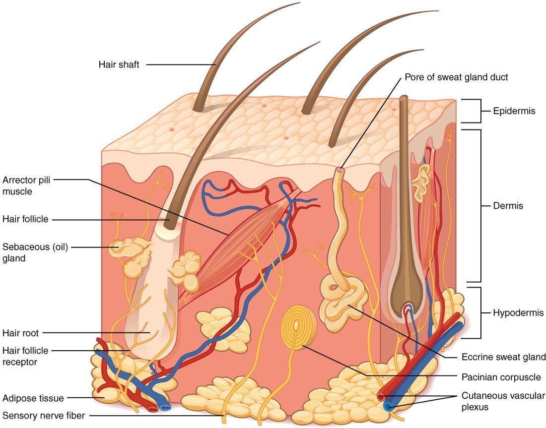
A skin section places epidermis above vascular dermis with follicles, glands, vessels, and sensory structures; subcutaneous tissue lies below. OpenStax College : J. Gordon Betts, Peter Desaix, Eddie Johnson.. 501 Structure of the skin.jpg. CC BY 3.0. Displayed at reduced size; anatomical labels unchanged.Skin and heat exchange. Skin blood flow and eccrine sweating are the main skin effectors for heat exchange. Their effect depends on environmental conditions, so a response does not guarantee adequate cooling or warming. Original diagram, McNova Enterprises, 2026; free to reuse in this course.Text description of this figure
The hypothalamus compares body temperature with its regulated range. Left branch, body temperature rises: dermal vessels dilate so more warm blood reaches the skin surface, and eccrine glands secrete sweat whose evaporation carries heat away; result, more heat lost from skin. Right branch, body temperature falls: dermal vessels constrict so heat is kept deeper, sweating decreases, and skeletal muscle shivering raises heat production; result, less heat lost and more produced. A note states that air temperature, humidity, clothing, and activity change how much heat these responses can move.
Lesson 5.1
Skin layers cells and appendages
Locate epidermis dermis and hypodermis
Relate keratinocytes melanocytes and glands to function
Distinguish thick from thin skin
Skin consists of epidermis and dermis. The hypodermis lies beneath and helps anchor skin; it is not one of skin's two layers. The epidermis is keratinized stratified squamous epithelium. Cells formed in the deeper strata move outward as they differentiate. Keratinized surface cells and extracellular lipids contribute to a barrier against water loss and external exposures.
The basal stratum contains proliferating keratinocytes and melanocytes. Melanocytes transfer melanin-containing structures to keratinocytes, helping protect nuclear material from ultraviolet exposure. Skin pigmentation reflects several factors, including melanin type and distribution; visible tone is not a measure of a person's health or identity. Epidermal immune and touch-associated cells add other functions.
The dermis has a superficial papillary region and a deeper reticular region containing connective tissue, vessels, nerves, and appendages. Thick skin on palms and soles has a prominent stratum corneum and a stratum lucidum, lacks hair follicles, and contains eccrine sweat glands. Hair-bearing thin skin covers most other regions. Sebaceous glands release lipid-rich material into follicles. Eccrine secretion supports cooling through evaporation. Apocrine glands have a different distribution and do not provide the main whole-body cooling pathway.
Apply the idea
Trace a pin from the skin surface through epidermis, dermis, and subcutaneous tissue on a diagram. State which layers contain blood vessels and why that matters for nutrition.
Compare your reasoning
The epidermis is avascular and obtains nutrients by diffusion from dermal vessels. Dermis and subcutaneous tissues contain blood vessels. Do not perform a puncture.
Thick skin has a prominent stratum corneum and a stratum lucidum, lacks hair follicles, and contains eccrine sweat glands.
Lesson 5.2
Barrier temperature and vitamin D
Explain skin contributions to water balance and heat exchange
Trace the start of vitamin D production
Predict effects of barrier damage
An intact epidermal barrier limits water loss and entry of many harmful substances. Skin also contributes to sensation and immune defense. Barrier damage can increase fluid loss and vulnerability to infection, while injury depth influences which vessels, receptors, and appendages remain. A visible surface area alone does not establish severity.
Skin blood flow changes heat transfer between internal tissues and the surface. Sweat evaporation removes heat when environmental conditions permit. Vasodilation can increase heat loss in a cooler environment, while vasoconstriction reduces it. In hot environments, heat exchange also depends on air temperature, humidity, clothing, and activity. Sweating is a physiological response, not a guarantee of adequate cooling.
Ultraviolet B exposure starts vitamin D synthesis in skin. Subsequent processing in the liver and kidneys produces the active hormonal form. This pathway connects skin with calcium regulation and bone. It does not mean unprotected ultraviolet exposure is necessary or appropriate for every person. Nutrition, pigmentation, geography, age, kidney function, and other factors influence the pathway. Explain the biological steps without turning them into individualized exposure or supplement advice.
Apply the idea
Build a three-system map linking skin, kidney, and bone through vitamin D and calcium. Add a separate heat-loss pathway.
Compare your reasoning
Skin begins vitamin D synthesis, the liver and kidney process it, and active vitamin D supports intestinal calcium absorption and bone mineral balance. Sweat evaporation and changing dermal blood flow are heat-exchange pathways.
UVB exposure starts vitamin D synthesis in skin. Later processing in the liver and kidneys produces the active hormonal form, which links skin to calcium regulation and bone.
Lesson 5.3
Skin injury and disease mechanisms
Compare partial and full thickness injury
Distinguish pressure injury from a thermal burn
Relate psoriasis and skin cancer to altered cellular behavior
A superficial burn involves epidermis. Partial thickness injury extends into dermis to varying depths, while full thickness injury destroys epidermis and dermis. Appearance, pain, and sensation vary with depth and location; absence of pain does not establish a minor injury because sensory structures may be damaged. This course uses layer-based models rather than asking learners to classify personal injuries.
Pressure injury arises from sustained tissue deformation and pressure, often with shear, and can involve deeper tissue before surface changes are obvious. Thermal burns and pressure injuries have different mechanisms and classification systems. A photo without clinical context is insufficient to stage a pressure injury or determine burn management. Across skin tones, assess more than redness: temperature, texture, pain, and other changes may matter clinically.
Psoriasis involves immune-mediated changes and rapid epidermal turnover. Skin cancers involve abnormal cell growth; basal cell, squamous cell, and melanoma arise from different cell populations. A changing pigmented lesion deserves clinical assessment, but a classroom image cannot establish its diagnosis. Compare mechanisms: barrier destruction, mechanical tissue injury, immune-mediated inflammation, and uncontrolled cellular growth are different starting points that may share visible consequences.
Apply the idea
For a fictional injury extending through epidermis into dermis, explain two functions that may be impaired. State why depth alone cannot determine the entire clinical risk.
Compare your reasoning
Barrier and sensory or temperature functions may be affected. Area, site, mechanism, age, associated injuries, and clinical condition also affect risk; depth alone is insufficient.
Pressure injury arises from sustained deformation and pressure, often with shear, and can involve deeper tissue before surface changes are obvious. It has a different mechanism and classification from burns.
Watch for these errors
Common error: The hypodermis is the third layer of the skin.
Better reasoning: Skin has two layers, epidermis and dermis. The hypodermis lies beneath and anchors skin but is not part of it.
Common error: Sweating guarantees that the body will cool down.
Better reasoning: Cooling needs evaporation. High humidity, clothing, and activity can limit heat loss even while sweat is produced.
Draw to learn: a section through skin
Producing your own labeled sketch from memory, then checking it, is a generative activity that supports later recall. Paper, a tablet, or McNova Drawing all work.
Draw a labeled cross-section of thin skin from the surface down to the hypodermis. Then mark how deep a superficial, a partial thickness, and a full thickness injury would reach.
Self-check after drawing
Virtual lab
Skin structure and injury mapping
Investigate How does modeled injury depth change the structures at risk?
Synthetic teaching data · Estimated work is included in this module laboratory allocation · 15 point report
Procedure
Use the skin illustration and supplied layer map.
For each model injury, shade only the named layers on a copy or describe them in text.
List two structures potentially affected.
Compare barrier continuity, appendage survival, and sensation as separate variables.
Explain why the model cannot determine treatment or clinical staging.
Model
Layers involved
Observation supplied
A
Epidermis only
Surface barrier disrupted
B
Epidermis and upper dermis
Some deeper appendages remain
C
Entire epidermis and dermis
Appendages within the injury destroyed
Analysis and submission
Which model preserves the most dermal structures?
Why can reduced sensation accompany greater depth?
Which information about extent is missing?
Submit an evidence sheet with a prediction, the provided observations, your graph or text equivalent, calculations with units, and a limitation. In public practice, save it on your own device.
Model limits These are synthetic layer maps. No burn images, skin self-examination, pressure application, or home injury experiments are required.
Expected interpretation and calculation check
A preserves dermal structures. C can destroy receptors and nerves in the affected tissue. Total involved area and body site are missing. B can retain some sources of epithelial regrowth, but healing depends on more than depth.
Clinical reasoning
A disrupted barrier
A fictional model has a broad loss of epidermal continuity. Its water loss increases.
Your task Explain the structure-function link and name one additional consequence that is plausible but unconfirmed.
Use structure → normal function → disruption → observable consequence → uncertainty. Suggested response 150 to 250 words; cases remain fictional.
Case discussion
Loss of the keratinized lipid-containing barrier permits greater water loss. Increased vulnerability to infection is plausible, but the case does not show an infection.
Explain it in your own words
Explain why a full thickness burn can be less painful than a partial thickness burn, and why that makes pain a poor guide to severity.
Compare with a model explanation
Pain depends on sensory receptors and nerve endings in the dermis. A partial thickness injury damages the dermis but leaves many receptors intact and irritated, so it can be very painful. A full thickness injury destroys the epidermis and dermis, including the receptors and nerves in that tissue, so the area may feel less pain. Reduced pain can mean deeper damage, not a minor injury.
Spaced review
These items return to earlier modules on purpose. Retrieving older material after a delay, mixed with new topics, strengthens long-term retention and helps you choose the right method when problem types are mixed.
Aligned collagen bundles suit loading mainly along their direction.
Oxygen supports electron transport; reduced oxidative ATP supply can limit ATP-dependent transport.
Close the module
1. Free recall (3 minutes, book closed). From memory, list the skin layers and the cell types in the epidermis, then trace the vitamin D pathway from skin to active hormone.
2. Rate your confidence on each objective (1 not yet, 4 confident), then take the checkpoint and compare. Large gaps between confidence and score show where to restudy.
Objective
1
2
3
4
Skin layers cells and appendages
Barrier temperature and vitamin D
Skin injury and disease mechanisms
3. Muddiest point. Write one sentence naming what is still least clear, and plan when you will revisit it (for example, in two days and again in a week).
Module checkpoint
Four questions worth 5 practice points each. Use the course player for saved attempts, or use the immediate practice feedback below.
Discussion or reflection
Choose one observation from this module. Explain it in 120 to 180 words using a mechanism, compare a plausible alternative, and state what evidence would distinguish them. In a group, respond to one peer using a source and a question. In public self-study, write the alternative yourself. No external account or public posting is required.
Relate bone structure and cells to remodeling, growth, fracture repair, and the landmarks used to orient the skeleton.
Suggested sequence (about 150 minutes of focused study, plus practice)
Make a prediction (2 minutes)
Lesson: Bone structure and remodeling, then its retrieval check
Lesson: Bone growth fracture healing and mineral balance, then its retrieval check
Lesson: Axial and appendicular anatomy, then its retrieval check
Watch for common errors and complete the drawing task
Virtual lab: Skeletal landmarks and model remodeling
Clinical reasoning case: A fracture after a low energy event
Explain it in your own words
Spaced review and close the module
Module checkpoint (four questions)
By the end you should be able to
Compare compact and spongy bone
Distinguish osteoblasts osteocytes and osteoclasts
Explain remodeling as coupled resorption and formation
Compare intramembranous and endochondral ossification
Describe growth plate activity
Explain fracture repair and osteoporosis
Locate major axial and appendicular bones
Identify functional bone markings
Orient a long bone from landmarks
Before you start: make a prediction
Guessing is expected here. Attempting a question before the lesson tends to make the explanation that follows easier to remember, even when the first answer is wrong. This item is not scored.
Bone is living connective tissue. Remodeling replaces existing bone through coordinated resorption by osteoclasts and formation by osteoblasts throughout life.
A long bone section locates shaft and ends, compact outer bone, internal spongy bone, marrow space, and surface coverings. OpenStax College. 603 Anatomy of Long Bone.jpg. CC BY 3.0. Displayed at reduced size; anatomical labels unchanged.The bone remodeling cycle. Remodeling replaces existing bone through coordinated resorption and formation. Loading, hormones such as parathyroid hormone, nutrition, and age change the balance between the two. Original diagram, McNova Enterprises, 2026; free to reuse in this course.Text description of this figure
Four boxes arranged in a clockwise cycle. 1, activation: osteocytes and hormones signal a site. 2, resorption: osteoclasts dissolve mineral and digest matrix. 3, reversal: the cavity is prepared and resorption stops. 4, formation: osteoblasts lay down osteoid that mineralizes. The cycle returns to activation. The center reads coupled resorption and formation, and notes that some osteoblasts become osteocytes in lacunae. A footnote states that if resorption exceeds formation over many cycles, bone mass and architecture decline.
Lesson 6.1
Bone structure and remodeling
Compare compact and spongy bone
Distinguish osteoblasts osteocytes and osteoclasts
Explain remodeling as coupled resorption and formation
Bone is living connective tissue with a matrix containing collagen and mineral. Collagen contributes tensile resistance, while mineral supports resistance to compression. Compact bone forms dense outer regions; spongy bone has trabeculae with spaces that can contain marrow. Spongy bone is not immature or weak bone by definition: trabecular organization supports loads with less mass.
In compact bone, osteons organize lamellae around central canals containing vessels and nerves. Osteocytes occupy lacunae and communicate through canaliculi. Osteoblasts form new matrix, while osteoclasts resorb bone. Osteocytes help detect mechanical conditions and coordinate signals. Periosteum covers most external surfaces, and endosteum lines internal surfaces. Articular cartilage covers joint surfaces rather than periosteum.
Remodeling replaces existing tissue through coordinated resorption and formation. Its balance reflects mechanical loading, hormones, nutrition, and age. Bone is a mineral reservoir as well as a structural tissue. Parathyroid hormone and active vitamin D participate in calcium regulation through interactions among bone, kidneys, and intestine. A fall in circulating calcium does not mean bone senses a command to release all its mineral at once; regulation involves receptors, cells, and time-dependent responses.
Apply the idea
Explain why a tissue can be mineralized yet remain metabolically active. Connect one microscopic feature to nutrient delivery.
Compare your reasoning
Bone contains living cells, vascular channels, and communicating processes. Central and perforating canals support vessels, while canaliculi support exchange around osteocytes.
Collagen contributes tensile resistance; mineral supports resistance to compression. Together they make bone strong without being brittle.
Lesson 6.2
Bone growth fracture healing and mineral balance
Compare intramembranous and endochondral ossification
Describe growth plate activity
Explain fracture repair and osteoporosis
Intramembranous ossification forms bone within a connective tissue membrane. Endochondral ossification replaces a cartilage model. Longitudinal growth of a long bone occurs through organized cartilage growth and replacement at the epiphyseal plate. Appositional growth adds tissue at surfaces and contributes to changes in diameter. After growth plates close, ordinary increases in adult height do not occur through renewed growth plate activity.
Fracture repair commonly includes hematoma formation, a fibrocartilaginous callus, a bony callus, and remodeling. These phases overlap. Stability, blood supply, infection, tissue damage, and individual factors affect healing. A callus bridges the injury before the final remodeled arrangement develops. A visible callus does not prove the original strength has returned.
Osteoporosis involves reduced bone strength and increased fracture susceptibility, reflecting bone mass and architecture. It can progress without symptoms until a fracture occurs. Osteomalacia involves impaired mineralization and is not a synonym for osteoporosis. Osteomyelitis is bone infection; Paget disease involves disordered remodeling. These conditions differ in mechanism and cannot be distinguished from a single classroom radiograph. Clinical review should preserve that distinction when connecting appearance to possible causes.
Apply the idea
Compare osteoporosis, osteomalacia, and osteomyelitis using one sentence per mechanism. Explain why one low-density image cannot distinguish them.
Compare your reasoning
Osteoporosis reduces strength through loss and architectural change; osteomalacia impairs mineralization; osteomyelitis is infection. Imaging must be interpreted with clinical and other evidence.
A callus bridges the injury before final remodeling. A visible callus does not prove that the original strength has returned.
Lesson 6.3
Axial and appendicular anatomy
Locate major axial and appendicular bones
Identify functional bone markings
Orient a long bone from landmarks
The axial skeleton includes skull, vertebral column, and thoracic cage. The appendicular skeleton includes limb girdles and limbs. The skull protects the brain and supports facial structures. Vertebrae surround the vertebral canal; cervical, thoracic, lumbar, sacral, and coccygeal regions differ in form. Ribs and sternum contribute to thoracic protection and movement during breathing.
The pectoral girdle consists of clavicles and scapulae. Upper limbs include humerus, radius, ulna, carpals, metacarpals, and phalanges. In anatomical position, radius is lateral to ulna. The pelvic girdle attaches lower limbs to the axial skeleton; each hip bone develops from ilium, ischium, and pubis. Lower limbs include femur, patella, tibia, fibula, tarsals, metatarsals, and phalanges. Tibia is medial to fibula.
Bone markings often reflect attachments, articulations, or passages. A tuberosity can provide an attachment area; a condyle forms part of an articular surface; a foramen permits passage. To orient a bone, use several landmarks together. On the femur, the head faces medially and the patellar surface faces anteriorly. A single broken or obscured landmark is weak evidence for assigning a side.
Apply the idea
Identify the sternum, scapula, humerus, radius, ulna, pelvis, femur, tibia, and fibula on the supplied or linked atlas. Write their system division and one relationship.
Compare your reasoning
Sternum is axial. Scapula and the named limb bones are appendicular. Radius is lateral to ulna; tibia is medial to fibula in anatomical position.
Orient a bone with several landmarks together. On the femur, the head faces medially and the patellar surface faces anteriorly. A single damaged landmark is weak evidence.
Watch for these errors
Common error: Spongy bone is weak or immature bone.
Better reasoning: Trabecular organization supports loads with less mass. Spongy bone is a normal, functional arrangement.
Common error: Osteoporosis and osteomalacia are the same condition.
Better reasoning: Osteoporosis is reduced bone strength from mass and architecture changes; osteomalacia is impaired mineralization. They differ in mechanism.
Draw to learn: an osteon and a long bone
Producing your own labeled sketch from memory, then checking it, is a generative activity that supports later recall. Paper, a tablet, or McNova Drawing all work.
Draw a long bone showing epiphyses, diaphysis, growth plate, compact and spongy bone, and periosteum. Add an enlarged osteon with a central canal, lamellae, lacunae, and canaliculi.
Self-check after drawing
Virtual lab
Skeletal landmarks and model remodeling
Investigate How do anatomical landmarks and formation-resorption balance support different kinds of conclusions?
Synthetic teaching data · Estimated work is included in this module laboratory allocation · 15 point report
Procedure
Locate ten bones using an illustrated atlas and record two landmarks for each.
Orient the humerus or femur from at least two landmarks; a text description is an acceptable alternative.
For the synthetic remodeling data, calculate formation minus resorption for each interval.
Graph the cumulative relative bone balance starting at zero.
Explain why balance units do not equal a clinical bone density measurement.
Interval
Formation units
Resorption units
1
5
4
2
5
6
3
4
7
4
4
6
Analysis and submission
What is the net change per interval?
What is cumulative balance after four intervals?
Why is fracture risk not determined by this number alone?
Submit an evidence sheet with a prediction, the provided observations, your graph or text equivalent, calculations with units, and a limitation. In public practice, save it on your own device.
Model limits Arbitrary model units do not represent a DXA result, a T score, or a patient trajectory. No fracture diagnosis is made.
Expected interpretation and calculation check
Net changes are +1, −1, −3, and −2; cumulative balance ends at −5 units. Architecture, tissue quality, loading, falls, and clinical factors also matter. Landmark evidence supports orientation, not disease diagnosis.
Clinical reasoning
A fracture after a low energy event
A fictional case describes a fracture after a fall from standing height, with no laboratory or density measurements supplied.
Your task Explain why bone strength is relevant and list evidence needed before attributing the fracture to osteoporosis.
Use structure → normal function → disruption → observable consequence → uncertainty. Suggested response 150 to 250 words; cases remain fictional.
Case discussion
Reduced bone strength is one possibility, but event mechanics, imaging, history, and clinical assessment are needed. A low energy fracture warrants evaluation; it does not establish a specific tissue mechanism from this paragraph alone.
Explain it in your own words
Explain how bone can lose mass over years even though resorption and formation both continue.
Compare with a model explanation
Remodeling couples resorption by osteoclasts with formation by osteoblasts at the same site. Mass is kept only if the amount formed matches the amount removed in each cycle. If loading falls, hormone levels change, or nutrition is poor, formation can fall slightly short of resorption. A small deficit repeated over many cycles produces a net loss of mass and changes in architecture, which reduces strength and raises fracture susceptibility without any symptoms until a fracture occurs.
Spaced review
These items return to earlier modules on purpose. Retrieving older material after a delay, mixed with new topics, strengthens long-term retention and helps you choose the right method when problem types are mixed.
Skin comprises epidermis and dermis, with hypodermis beneath.
DNA replication precedes mitosis and occurs during S phase of interphase.
Close the module
1. Free recall (3 minutes, book closed). List the three bone cell types with their roles, the four stages of fracture repair, and the bones of the upper and lower limbs.
2. Rate your confidence on each objective (1 not yet, 4 confident), then take the checkpoint and compare. Large gaps between confidence and score show where to restudy.
Objective
1
2
3
4
Bone structure and remodeling
Bone growth fracture healing and mineral balance
Axial and appendicular anatomy
3. Muddiest point. Write one sentence naming what is still least clear, and plan when you will revisit it (for example, in two days and again in a week).
Module checkpoint
Four questions worth 5 practice points each. Use the course player for saved attempts, or use the immediate practice feedback below.
Discussion or reflection
Choose one observation from this module. Explain it in 120 to 180 words using a mechanism, compare a plausible alternative, and state what evidence would distinguish them. In a group, respond to one peer using a source and a question. In public self-study, write the alternative yourself. No external account or public posting is required.
Classify joints, describe movement with planes and axes, calculate simple torque, and compare mechanisms of joint injury and arthritis.
Suggested sequence (about 150 minutes of focused study, plus practice)
Make a prediction (2 minutes)
Lesson: Joint structure and classification, then its retrieval check
Lesson: Motion planes and mechanical advantage, then its retrieval check
Lesson: Joint injury osteoarthritis and inflammatory arthritis, then its retrieval check
Watch for common errors and complete the drawing task
Virtual lab: Joint angle and torque analysis
Clinical reasoning case: Two stiff joints
Explain it in your own words
Spaced review and close the module
Module checkpoint (four questions)
By the end you should be able to
Compare fibrous cartilaginous and synovial joints
Identify synovial joint components
Distinguish structural and functional classifications
Apply flexion extension abduction adduction and rotation
Relate movement to a plane and axis
Calculate a simplified joint torque
Distinguish sprain strain and dislocation
Compare osteoarthritis and rheumatoid arthritis mechanisms
Explain why symptoms alone do not establish a diagnosis
Before you start: make a prediction
Guessing is expected here. Attempting a question before the lesson tends to make the explanation that follows easier to remember, even when the first answer is wrong. This item is not scored.
Torque is force times moment arm. The biceps attaches close to the elbow, so its short moment arm requires a much larger force to balance the load held far out in the hand.
A synovial joint section shows opposing cartilage surfaces, a cavity containing fluid, and an enclosing capsule. OpenStax College. 907 Synovial Joints.jpg. CC BY 3.0. Displayed at reduced size; anatomical labels unchanged.Lever classes in the body. The class depends on where the fulcrum (joint), effort (muscle pull), and load sit along the lever. In class 3 levers the muscle works at a short moment arm, which is why muscle force can exceed the load held in the hand. Original diagram, McNova Enterprises, 2026; free to reuse in this course.Text description of this figure
Three lever diagrams stacked vertically. Class 1: the fulcrum lies between effort and load, as in head nodding at the atlanto-occipital joint, where neck extensors pull behind the joint and head weight acts in front of it. Class 2: the load lies between fulcrum and effort, as in rising onto the balls of the feet, where the ball of the foot is the fulcrum and calf muscles pull on the calcaneus. Class 3: the effort lies between fulcrum and load, as in elbow flexion by biceps brachii, the most common arrangement in the body; with a short effort arm, muscle force exceeds the hand load, but the hand moves farther and faster. A footnote gives torque equals force times perpendicular moment arm, and effort times effort arm equals load times load arm at balance.
Lesson 7.1
Joint structure and classification
Compare fibrous cartilaginous and synovial joints
Identify synovial joint components
Distinguish structural and functional classifications
A joint is a place where skeletal elements meet. Structural classification describes the connecting material and whether a joint cavity is present. Fibrous joints join elements through fibrous connective tissue; cartilaginous joints join them through cartilage. Synovial joints have an enclosed cavity. Functional classification describes movement: synarthrosis has little or no movement, amphiarthrosis permits limited movement, and diarthrosis is freely movable. These naming systems answer different questions.
A synovial joint includes articular cartilage on opposing surfaces, a capsule, a synovial membrane, and synovial fluid. Hyaline articular cartilage reduces friction and distributes load. Synovial fluid supports lubrication and nutrient exchange for cartilage. The fibrous capsule and ligaments constrain movement, while surrounding muscles contribute active stability. Some joints also contain discs, menisci, bursae, or tendon sheaths.
A ligament commonly connects bone to bone, while a tendon commonly transmits force from muscle to bone. Both contain collagen-rich connective tissue, but their roles are not identical. A stable joint balances congruence, passive constraints, and muscle control. Greater range of motion can require different stability arrangements. The shoulder's mobility and the hip's deeper socket illustrate why no single joint design fits every task.
Apply the idea
Draw a synovial joint and label cartilage, capsule, cavity, synovial lining, and a ligament. Describe one source of passive and one source of active stability.
Compare your reasoning
Capsule, ligament, and articular congruence contribute passive constraints; muscles contribute active stabilization. Synovial membrane lines internal noncartilaginous surfaces.
Functional classification describes movement: synarthrosis little or none, amphiarthrosis limited, diarthrosis free. Structural classification describes the connecting material and cavity. The two systems answer different questions.
Lesson 7.2
Motion planes and mechanical advantage
Apply flexion extension abduction adduction and rotation
Relate movement to a plane and axis
Calculate a simplified joint torque
Flexion usually decreases a joint angle and extension increases it. Abduction moves away from the reference midline, while adduction moves toward it. Internal and external rotation turn a structure around its long axis. Pronation and supination describe forearm movements; dorsiflexion and plantar flexion describe ankle motion. Always state the joint and reference position because the same everyday word can hide different anatomical actions.
Motion in one plane occurs around an axis perpendicular to that plane. Flexion and extension are commonly described in the sagittal plane around a mediolateral axis. Abduction and adduction commonly occur in a frontal plane around an anteroposterior axis. Many activities combine planes, and joint surfaces can translate as they rotate.
Torque equals force multiplied by its perpendicular moment arm. In a simplified static forearm model, a 20 N load at 0.30 m produces 6 N·m of external torque. A muscle with a 0.03 m perpendicular moment arm would need 200 N to balance that torque if other forces are ignored. Muscle force can exceed the hand load because its moment arm is shorter. Real joints involve multiple muscles, segment weights, changing geometry, and three-dimensional forces.
Apply the idea
Calculate torque for a 15 N load at 0.20 m and the balancing muscle force for a 0.03 m moment arm. State two omitted forces.
Compare your reasoning
External torque is 3 N·m; balancing force is 100 N. The model omits forearm weight, other muscle forces, and joint reaction forces.
External torque is 30 N × 0.25 m = 7.5 N·m. Dividing by the muscle's 0.05 m moment arm gives 7.5 / 0.05 = 150 N.
Lesson 7.3
Joint injury osteoarthritis and inflammatory arthritis
Distinguish sprain strain and dislocation
Compare osteoarthritis and rheumatoid arthritis mechanisms
Explain why symptoms alone do not establish a diagnosis
A sprain involves ligament injury; a strain involves muscle or tendon injury. A dislocation disrupts normal alignment of articular surfaces. Pain, swelling, and reduced movement can occur with several injuries. The name of the symptom does not establish the tissue involved, and trying to reproduce painful movement is not a course activity.
Osteoarthritis affects cartilage and other joint tissues, including bone and synovium. It is not explained adequately as passive wear alone. Altered tissue behavior can change load distribution and movement. Rheumatoid arthritis is an autoimmune inflammatory disease that often involves synovium and can damage cartilage and bone. It can also affect structures outside joints. These descriptions compare mechanisms rather than provide rules for diagnosing an individual.
Reduced motion may reflect pain, effusion, altered tissue structure, muscle guarding, or neurologic changes. A goniometer measurement describes an angle; it does not explain the cause of a limit. Use the same landmarks, starting position, and movement definition when comparing measurements. Measurement variation should be considered before treating a small difference as a biological change.
Apply the idea
Compare an angle measurement with a diagnostic conclusion. Explain why a smaller measured range could have more than one cause.
Compare your reasoning
An angle is an observation tied to a protocol. Pain, swelling, stiffness, guarding, and measurement technique can change it; further evidence is needed to attribute a cause.
Osteoarthritis affects cartilage, bone, and synovium and is not explained by wear alone. Rheumatoid arthritis is autoimmune and inflammatory, often involves synovium, and can affect structures outside joints.
Watch for these errors
Common error: A muscle only needs to pull as hard as the load it lifts.
Better reasoning: Most limb muscles work at a short moment arm, so they must produce much more force than the load. They gain speed and range of motion in return.
Common error: Sprain and strain are two words for the same injury.
Better reasoning: A sprain involves a ligament; a strain involves muscle or tendon. The symptom name does not identify the tissue.
Draw to learn: a synovial joint and a forearm lever
Producing your own labeled sketch from memory, then checking it, is a generative activity that supports later recall. Paper, a tablet, or McNova Drawing all work.
Draw a synovial joint and label articular cartilage, capsule, synovial membrane, synovial fluid, and a ligament. Then sketch the forearm as a lever and mark the fulcrum, effort, load, and both moment arms.
Self-check after drawing
Virtual lab
Joint angle and torque analysis
Investigate How do measurement variation and moment arm change a movement analysis?
Synthetic teaching data · Estimated work is included in this module laboratory allocation · 15 point report
Procedure
Use the three supplied readings rather than measuring yourself or another person.
Calculate the mean and range for each model joint angle.
Calculate external torque using the listed load and perpendicular distance.
Compare the two muscle moment arms while keeping external torque constant.
Report units and one source of measurement error.
Trial
Model A angle degrees
Model B angle degrees
1
118
102
2
120
105
3
119
104
Analysis and submission
Calculate both mean angles.
For 20 N at 0.25 m calculate external torque.
Compare muscle force at moment arms 0.025 m and 0.050 m.
Submit an evidence sheet with a prediction, the provided observations, your graph or text equivalent, calculations with units, and a limitation. In public practice, save it on your own device.
Model limits Synthetic angles are not reference ranges or impairment ratings. The torque model omits segment weights and cocontraction. No forced range-of-motion testing is needed.
Expected interpretation and calculation check
Mean A is 119°; mean B is about 103.7°. External torque is 5 N·m. Required simplified balancing forces are 200 N and 100 N. The longer moment arm requires less force for the same torque.
Clinical reasoning
Two stiff joints
One fictional joint has altered cartilage and subchondral bone. Another has persistent immune-mediated synovial inflammation.
Your task Compare the starting tissues and predict a shared functional consequence.
Use structure → normal function → disruption → observable consequence → uncertainty. Suggested response 150 to 250 words; cases remain fictional.
Case discussion
The first description is consistent with an osteoarthritis mechanism and the second with an inflammatory arthritis mechanism. Both can reduce comfortable movement. A symptom comparison alone cannot establish either diagnosis.
Explain it in your own words
Two people have the same reduced knee range measured with a goniometer. Explain why the measurement alone cannot tell you the cause.
Compare with a model explanation
A goniometer records an angle, not a mechanism. Reduced motion could come from pain, fluid in the joint, changes in cartilage or capsule structure, muscle guarding, or a neurologic change. Two people can share the same number for different reasons. Measurement method also matters: landmarks, starting position, and movement definition must match before a small difference is treated as a true change. Other evidence is needed to separate the possibilities.
Spaced review
These items return to earlier modules on purpose. Retrieving older material after a delay, mixed with new topics, strengthens long-term retention and helps you choose the right method when problem types are mixed.
These terms name different mechanisms and should not be used interchangeably.
Aligned collagen bundles suit loading mainly along their direction.
Close the module
1. Free recall (3 minutes, book closed). List the three structural and three functional joint classes, six movement terms with their planes, and the torque equation.
2. Rate your confidence on each objective (1 not yet, 4 confident), then take the checkpoint and compare. Large gaps between confidence and score show where to restudy.
Objective
1
2
3
4
Joint structure and classification
Motion planes and mechanical advantage
Joint injury osteoarthritis and inflammatory arthritis
3. Muddiest point. Write one sentence naming what is still least clear, and plan when you will revisit it (for example, in two days and again in a week).
Module checkpoint
Four questions worth 5 practice points each. Use the course player for saved attempts, or use the immediate practice feedback below.
Discussion or reflection
Choose one observation from this module. Explain it in 120 to 180 words using a mechanism, compare a plausible alternative, and state what evidence would distinguish them. In a group, respond to one peer using a source and a question. In public self-study, write the alternative yourself. No external account or public posting is required.
Trace the chain from motor neuron signal to cross-bridge force, and explain how whole muscles grade force and differ by type.
Suggested sequence (about 150 minutes of focused study, plus practice)
Make a prediction (2 minutes)
Lesson: Skeletal muscle organization and the neuromuscular junction, then its retrieval check
Lesson: Cross bridges recruitment and muscle metabolism, then its retrieval check
Lesson: Major muscles and tissue differences, then its retrieval check
Watch for common errors and complete the drawing task
Virtual lab: Recruitment and fatigue model
Clinical reasoning case: A signal with less force
Explain it in your own words
Spaced review and close the module
Module checkpoint (four questions)
By the end you should be able to
Order muscle fascicle fiber myofibril and sarcomere
Trace a neuromuscular signal
Explain excitation contraction coupling
Explain ATP roles in contraction and relaxation
Compare twitch summation and recruitment
Distinguish isometric from isotonic contraction
Relate muscle position to common actions
Compare skeletal cardiac and smooth muscle
Connect three muscle disorders to different mechanisms
Before you start: make a prediction
Guessing is expected here. Attempting a question before the lesson tends to make the explanation that follows easier to remember, even when the first answer is wrong. This item is not scored.
In the sliding filament model, thin and thick filaments slide past one another. The sarcomere shortens as Z discs move closer, but the filaments themselves do not shorten.
A skeletal muscle fiber contains myofibrils with repeating sarcomeres organized from thick myosin and thin actin filaments. OpenStax. 1002 Organization of Muscle Fiber.jpg. CC BY 4.0. Displayed at reduced size; anatomical labels unchanged.The cross-bridge cycle. Each cycle moves the thin filament a short distance toward the sarcomere center. ATP is needed for myosin to detach and to re-energize the head, and for calcium pumps during relaxation. Original diagram, McNova Enterprises, 2026; free to reuse in this course.Text description of this figure
Four boxes in a clockwise cycle. 1, attach: energized myosin holding ADP and phosphate binds an exposed actin site. 2, power stroke: phosphate is released, the head pivots, the thin filament slides, and ADP leaves. 3, detach: ATP binds myosin and the head releases actin. 4, re-energize: ATP is hydrolyzed to ADP and phosphate and the head is re-cocked, returning to step 1. The center states the cycle continues while calcium is bound to troponin and tropomyosin leaves binding sites exposed, and that without ATP myosin cannot detach, called rigor. A footnote says relaxation also uses ATP because pumps return calcium to the sarcoplasmic reticulum.
Lesson 8.1
Skeletal muscle organization and the neuromuscular junction
Order muscle fascicle fiber myofibril and sarcomere
Trace a neuromuscular signal
Explain excitation contraction coupling
A skeletal muscle contains fascicles, each containing muscle fibers. A fiber is one long cell containing myofibrils. Myofibrils contain repeating sarcomeres organized from thin and thick filaments. Endomysium, perimysium, and epimysium are connective tissue layers that support organization and transmission of force. Tendons link that force to skeletal attachments.
At the neuromuscular junction, a motor neuron action potential reaches its terminal and opens voltage-gated calcium channels. Calcium entry supports acetylcholine release. Acetylcholine binds nicotinic receptors on the motor end plate, producing a graded depolarization. If the adjacent membrane reaches threshold, a muscle action potential propagates. Acetylcholinesterase breaks down acetylcholine, helping terminate the signal.
The muscle action potential travels along sarcolemma and T tubules. This electrical event is coupled to calcium release from the sarcoplasmic reticulum. Calcium binds troponin, moving tropomyosin away from actin's binding sites. Myosin can then interact cyclically with actin. The motor end plate potential, muscle action potential, calcium release, and mechanical force are related but different events. A failure at one step can reduce force even if another step remains intact.
Apply the idea
Write a six-arrow pathway from motor neuron firing to calcium binding troponin. Mark where extracellular calcium and stored intracellular calcium have different roles.
Compare your reasoning
Terminal calcium entry triggers transmitter release. Muscle membrane excitation triggers release from sarcoplasmic reticulum; that calcium binds troponin. These are different calcium compartments and events.
Acetylcholinesterase breaks down acetylcholine and helps end the signal. Blocking it leaves acetylcholine in the cleft longer; release and calcium entry are earlier steps that are unaffected.
Lesson 8.2
Cross bridges recruitment and muscle metabolism
Explain ATP roles in contraction and relaxation
Compare twitch summation and recruitment
Distinguish isometric from isotonic contraction
During cross-bridge cycling, myosin binds actin, produces a power stroke, releases after ATP binds, and is reenergized through ATP hydrolysis. Thin and thick filaments slide past one another; they do not shorten themselves. Sarcomere length decreases as Z discs approach, while the A band remains approximately constant. Calcium removal by ATP-dependent pumps supports relaxation. ATP shortage can impair detachment and calcium removal, not simply reduce the number of power strokes.
A motor unit consists of one motor neuron and the skeletal muscle fibers it innervates. Recruitment activates additional motor units. Increasing firing frequency can produce temporal summation because force from individual responses overlaps. A whole muscle grades force through both mechanisms. An action potential in an excitable fiber is all-or-none in the usual model, while whole-muscle force is graded.
An isometric contraction develops force without a net change in whole-muscle length. In concentric action the muscle shortens while producing force; in eccentric action it lengthens under load. ATP supply comes from existing ATP, phosphocreatine, glycolysis, and oxidative metabolism in proportions that vary with activity. Fatigue has several mechanisms. Lactate is a metabolic product and fuel substrate, not a complete explanation of all fatigue or delayed soreness.
Apply the idea
Explain why holding an object still can require ATP. Predict how recruitment can increase force without increasing the amplitude of each muscle fiber action potential.
Compare your reasoning
Cross-bridge activity, calcium handling, and ion gradients use energy during a hold. Recruiting more motor units adds active fibers; it does not require larger individual action potentials.
A single fiber's action potential is all-or-none, but whole-muscle force is graded through recruitment of more motor units and temporal summation from higher firing frequency.
Lesson 8.3
Major muscles and tissue differences
Relate muscle position to common actions
Compare skeletal cardiac and smooth muscle
Connect three muscle disorders to different mechanisms
Learn muscle actions from attachments and the joint crossed, then verify them in an atlas. Deltoid contributes to shoulder abduction. Biceps brachii contributes to elbow flexion and forearm supination; triceps brachii extends the elbow. Quadriceps extend the knee, hamstrings flex it, and gastrocnemius contributes to plantar flexion and knee flexion. Tibialis anterior dorsiflexes the ankle. Gluteus maximus contributes to hip extension. Individual muscles often have several actions depending on position.
An agonist contributes to a selected movement, an antagonist opposes that action, and synergists assist or stabilize. These are task-dependent roles. During controlled lowering, a muscle can generate force while lengthening. Surface location alone does not establish the direction of force or whether a muscle is active.
Cardiac muscle is striated and linked through intercalated discs. Smooth muscle lacks sarcomeric striation and regulates contraction through calcium-calmodulin and myosin light-chain phosphorylation rather than troponin. Myasthenia gravis involves impaired neuromuscular transmission, often associated with antibodies affecting postsynaptic structures. Muscular dystrophies involve inherited changes affecting muscle integrity. Rhabdomyolysis involves breakdown of skeletal muscle with release of cellular contents; dark urine and muscle pain are not a complete diagnostic definition. The distinctions locate the disrupted process without asking learners to select treatment.
Apply the idea
For elbow flexion and controlled lowering, describe biceps role, direction of length change, and need for energy. Compare junction failure with damage to the muscle fiber itself.
Compare your reasoning
Biceps can shorten during flexion and lengthen while controlling extension. Both require active force and energy. Junction dysfunction can limit activation even when contractile proteins remain present.
Smooth muscle lacks sarcomeric striation and regulates contraction through calcium-calmodulin and myosin light-chain phosphorylation rather than the troponin system.
Watch for these errors
Common error: Myosin heads detach from actin because the power stroke is finished.
Better reasoning: Detachment requires a new ATP to bind myosin. Without ATP, heads stay attached, which is the basis of rigor.
Common error: Lactate build-up explains all muscle fatigue and soreness.
Better reasoning: Fatigue has several mechanisms, and lactate is also a fuel. It does not explain all fatigue or delayed soreness.
Draw to learn: a sarcomere before and after contraction
Producing your own labeled sketch from memory, then checking it, is a generative activity that supports later recall. Paper, a tablet, or McNova Drawing all work.
Draw one relaxed and one contracted sarcomere. Label Z discs, thin filaments, thick filaments, the A band, the I band, and the H zone in both.
Self-check after drawing
Virtual lab
Recruitment and fatigue model
Investigate How does motor unit activation change modeled force, and how can repeated activation alter output?
Synthetic teaching data · Estimated work is included in this module laboratory allocation · 15 point report
Procedure
Use only model data; no electrical stimulation or sustained exercise is required.
Plot active motor units against force.
Calculate force per active unit in the recruitment series.
Compare a separate repeated-stimulation series with outputs 10, 9, 8, 7, and 6 arbitrary units.
Explain why neither series alone measures an individual fiber action potential amplitude.
Active model units
Force arbitrary units
1
2
2
4
3
6
4
8
5
10
Analysis and submission
Does this simplified model add units linearly?
What is the percent force reduction from 10 to 6?
Name two physiological details omitted.
Submit an evidence sheet with a prediction, the provided observations, your graph or text equivalent, calculations with units, and a limitation. In public practice, save it on your own device.
Model limits The linear model is illustrative and is not EMG. A clinical EMG records electrical activity using an appropriate instrument and interpretation. No electrodes, current, or fatigue challenge are needed.
Expected interpretation and calculation check
Force rises by 2 units per active model unit. The repeated series falls 40%. Real units differ in size and recruitment thresholds; firing rate, length, and metabolic conditions also affect force.
Motor unit recruitment model
Each model unit contributes 2 force units. Real motor units differ in size, and firing rate and muscle length also affect force. This is not an EMG recording.
Clinical reasoning
A signal with less force
A fictional neuromuscular model shows normal motor axon impulses but reduced activation of muscle fibers.
Your task Locate a plausible failure between nerve terminal and muscle excitation. Name an alternative downstream explanation.
Use structure → normal function → disruption → observable consequence → uncertainty. Suggested response 150 to 250 words; cases remain fictional.
Case discussion
Transmitter release, receptor function, or end plate depolarization could be altered. If excitation is present, calcium handling or contractile function could instead explain reduced force. The pattern is not enough to diagnose myasthenia gravis.
Explain it in your own words
A fictional record shows normal motor nerve signals but weak muscle force. Explain three places in the excitation-contraction chain where the problem could lie.
Compare with a model explanation
First, transmission at the neuromuscular junction could fail: less acetylcholine release or fewer working receptors would give an end plate potential that does not reach threshold. Second, the muscle action potential might not trigger enough calcium release from the sarcoplasmic reticulum. Third, cross-bridge cycling might be impaired, such as by low ATP or damaged contractile proteins. Each step is a different event, so the same weak force can have different starting points.
Spaced review
These items return to earlier modules on purpose. Retrieving older material after a delay, mixed with new topics, strengthens long-term retention and helps you choose the right method when problem types are mixed.
A cavity within a capsule distinguishes synovial joints structurally.
Skin comprises epidermis and dermis, with hypodermis beneath.
Close the module
1. Free recall (3 minutes, book closed). Order the steps from motor neuron action potential to cross-bridge cycling, then compare skeletal, cardiac, and smooth muscle in three features each.
2. Rate your confidence on each objective (1 not yet, 4 confident), then take the checkpoint and compare. Large gaps between confidence and score show where to restudy.
Objective
1
2
3
4
Skeletal muscle organization and the neuromuscular junction
Cross bridges recruitment and muscle metabolism
Major muscles and tissue differences
3. Muddiest point. Write one sentence naming what is still least clear, and plan when you will revisit it (for example, in two days and again in a week).
Module checkpoint
Four questions worth 5 practice points each. Use the course player for saved attempts, or use the immediate practice feedback below.
Discussion or reflection
Choose one observation from this module. Explain it in 120 to 180 words using a mechanism, compare a plausible alternative, and state what evidence would distinguish them. In a group, respond to one peer using a source and a question. In public self-study, write the alternative yourself. No external account or public posting is required.
Follow a neural signal from resting membrane voltage through action potentials and synapses to brain, spinal, reflex, and autonomic pathways.
Suggested sequence (about 300 minutes of focused study, plus practice)
Make a prediction (2 minutes)
Lesson: Neurons glia and resting membrane potential, then its retrieval check
Lesson: Graded potentials and action potentials, then its retrieval check
Lesson: Synapses and pathway failure, then its retrieval check
Lesson: Brain spinal cord and protective structures, then its retrieval check
Lesson: Peripheral nerves cranial nerves and reflexes, then its retrieval check
Lesson: Autonomic control and neural disease localization, then its retrieval check
Watch for common errors and complete the drawing task
Virtual lab: Conduction time and signal interpretation
Clinical reasoning case: Slower propagation
Explain it in your own words
Spaced review and close the module
Module checkpoint (four questions)
By the end you should be able to
Identify soma dendrites axon and terminals
Compare central and peripheral myelination
Explain selective permeability and resting voltage
Distinguish graded from all-or-none signals
Trace sodium and potassium channel events
Explain refractory periods and saltatory conduction
Trace chemical synaptic transmission
Compare excitatory and inhibitory postsynaptic effects
Distinguish conduction failure from loss of transmitter response
Locate major brain regions
Compare gray and white matter
Trace cerebrospinal fluid flow at an introductory level
Distinguish nerve tract ganglion and nucleus
Identify the twelve cranial nerves and selected functions
Trace a reflex arc
Compare somatic and autonomic motor pathways
Explain sympathetic and parasympathetic organization
Use a pathway map without overdiagnosing
Before you start: make a prediction
Guessing is expected here. Attempting a question before the lesson tends to make the explanation that follows easier to remember, even when the first answer is wrong. This item is not scored.
Action potential amplitude is relatively fixed in a given axon. Stimulus strength is represented by firing pattern and by recruiting more pathways, not by taller spikes.
A nervous system overview distinguishes brain and spinal cord from peripheral nerves extending to the body. OpenStax. 1201 Overview of Nervous System.jpg. CC BY 4.0. Displayed at reduced size; anatomical labels unchanged.A brainstem illustration locates midbrain, pons, and medulla in relation to adjacent brain regions. OpenStax. 1311 Brain Stem.jpg. CC BY 4.0. Displayed at reduced size; anatomical labels unchanged.Phases of an action potential. Teaching values from a standard axon model: resting near −70 mV, threshold near −55 mV, and a peak near +30 mV. Individual neurons vary, and the exact timing depends on the axon and temperature. Original diagram, McNova Enterprises, 2026; free to reuse in this course.Text description of this figure
A graph of membrane voltage in millivolts against time in milliseconds from 0 to 5. The trace begins flat at about minus 70 millivolts, the resting value. It rises slowly to a dashed threshold line at about minus 55, then climbs steeply during depolarization as sodium channels open, peaking at about plus 30 millivolts near 1.5 milliseconds, where sodium channels inactivate. It falls during repolarization as potassium channels open and potassium leaves, dips below rest to about minus 80 during afterhyperpolarization because potassium channels are slow to close, and returns to minus 70 by about 4.5 milliseconds. Bars beneath the time axis mark the absolute refractory period, from threshold through most of repolarization, followed by the relative refractory period during afterhyperpolarization, when a stronger input is needed.
The spinal reflex arc. A stretch reflex shown as five linked parts. Mapping a deficit to one part generates a hypothesis; it does not identify a cause on its own. Original diagram, McNova Enterprises, 2026; free to reuse in this course.Text description of this figure
Five boxes in a loop. 1, receptor: a muscle spindle detects stretch. An afferent signal travels to 2, afferent neuron, which enters the spinal cord by the dorsal root and has its cell body in the dorsal root ganglion. It reaches 3, integration: a synapse in spinal cord gray matter. Then 4, efferent neuron: a motor neuron leaves by the ventral root. An efferent signal reaches 5, effector: skeletal muscle contracts. A note states that a missing response could involve any of the five parts or the testing technique, and that descending pathways from the brain can modify the response without a conscious decision being needed.
Lesson 9.1
Neurons glia and resting membrane potential
Identify soma dendrites axon and terminals
Compare central and peripheral myelination
Explain selective permeability and resting voltage
A typical neuron has a cell body, dendrites, an axon, and terminals, although shapes vary with function. Dendrites and the soma often receive signals; an axon propagates action potentials toward terminals. Sensory neurons, interneurons, and motor neurons occupy different roles in information pathways. Directional terms such as afferent and efferent are defined relative to the stated reference, commonly the central nervous system.
Glia support chemical balance, insulation, defense, and other functions. Oligodendrocytes form myelin around central axons, while Schwann cells myelinate peripheral axons. Astrocytes support the neural environment and blood-brain barrier functions in cooperation with other cells. Microglia participate in immune surveillance. Myelin reduces current loss and changes how depolarization spreads between nodes of Ranvier.
Resting membrane voltage results from ion concentration gradients and selective permeability, especially potassium permeability in many neurons. The sodium potassium pump maintains gradients over time and also has an electrogenic contribution. It is misleading to attribute the entire resting voltage only to the pump. A value near −70 mV is a common teaching example, not a universal voltage for every neuron or every moment.
Apply the idea
Compare the role of ion channels with the sodium potassium pump in resting voltage. Identify one CNS and one PNS myelinating cell.
Resting voltage results from ion gradients and selective permeability, especially to potassium. The pump maintains gradients and adds a small electrogenic part, but it is misleading to credit the whole voltage to the pump.
Lesson 9.2
Graded potentials and action potentials
Distinguish graded from all-or-none signals
Trace sodium and potassium channel events
Explain refractory periods and saltatory conduction
Graded potentials vary in size, can be depolarizing or hyperpolarizing, and generally diminish with distance. Their spatial and temporal summation influences whether an excitable region reaches threshold. An action potential is a regenerative event whose amplitude is relatively stereotyped in a particular axon under given conditions. A stronger sensory input is commonly represented by firing pattern and recruited pathways rather than unlimited action potential height.
In a standard axon model, voltage-gated sodium channels open rapidly during depolarization and then inactivate. Delayed potassium conductance contributes to repolarization and afterhyperpolarization. Channel inactivation and recovery help produce refractory periods. The absolute refractory period prevents another action potential during part of the cycle; the relative refractory period requires a stronger depolarizing input.
Local current brings adjacent membrane toward threshold. In myelinated axons, regenerative events occur at nodes, producing saltatory conduction. Signals do not literally leap through empty space; current spreads through insulated segments and activates the next node. Conduction velocity depends on properties including diameter, myelination, and temperature. Demyelination may slow conduction or lead to failure without necessarily destroying every affected axon.
Apply the idea
Sketch a labeled action potential and describe which channel behavior supports each phase. Explain why increasing stimulus strength usually does not double action potential height.
Compare your reasoning
Rapid sodium conductance drives the upstroke; inactivation and delayed potassium conductance support recovery. Thresholded regenerative behavior limits amplitude variation; stimulus strength is commonly encoded in frequency and recruitment.
In the absolute refractory period another action potential cannot be produced. In the relative refractory period, a stronger depolarizing input can still reach threshold.
Lesson 9.3
Synapses and pathway failure
Trace chemical synaptic transmission
Compare excitatory and inhibitory postsynaptic effects
Distinguish conduction failure from loss of transmitter response
At a chemical synapse, an arriving action potential opens terminal calcium channels. Calcium-dependent vesicle fusion releases transmitter into a cleft. The transmitter binds postsynaptic receptors and changes activity through ion channels or intracellular pathways. Termination can involve reuptake, enzymatic breakdown, or diffusion. The presynaptic action potential and postsynaptic response are separate events.
An excitatory postsynaptic potential increases the likelihood of reaching threshold under the relevant conditions; an inhibitory effect reduces that likelihood. The transmitter name alone does not always determine the effect because receptor type and ion gradients matter. The same signaling molecule can act differently in different tissues. Electrical synapses permit current through gap junctions and differ from this vesicle-mediated sequence.
Multiple sclerosis involves immune-mediated damage in the central nervous system, including myelin and, in some lesions, axons. It is not a general disease of every peripheral Schwann cell. Parkinson disease is associated with loss of dopaminergic neurons in the substantia nigra and altered movement circuits. Alzheimer disease involves progressive changes affecting cognition and neural networks. These examples locate different disruptions; similar functional complaints can arise through different mechanisms and require clinical evaluation.
Apply the idea
A model axon conducts normally but its postsynaptic cell does not respond. List two possible synaptic failures and one experiment that would distinguish them.
Compare your reasoning
Reduced transmitter release and impaired postsynaptic receptor response are possible. Directly applying transmitter in a controlled model could test postsynaptic responsiveness, but it would not alone characterize every downstream pathway.
The transmitter name alone does not always determine the effect. Receptor type and the ion gradients across the postsynaptic membrane set whether the response is excitatory or inhibitory.
Lesson 9.4
Brain spinal cord and protective structures
Locate major brain regions
Compare gray and white matter
Trace cerebrospinal fluid flow at an introductory level
The cerebrum supports perception, voluntary movement, language, memory, and other functions through interacting networks. The diencephalon includes thalamic and hypothalamic regions. The brainstem includes midbrain, pons, and medulla. The cerebellum contributes to coordination, timing, and error correction in movement. Functions overlap across networks; a lobe name is not a complete explanation of a person's abilities.
Gray matter contains many neuronal cell bodies, dendrites, and synapses. White matter contains many myelinated axons. In the spinal cord, central gray matter forms dorsal and ventral horns, while white matter pathways surround it. Dorsal roots carry afferent fibers and contain sensory neuron cell bodies in dorsal root ganglia. Ventral roots carry efferent fibers. Once roots join, a typical spinal nerve contains mixed fibers.
Meninges include dura mater, arachnoid mater, and pia mater. Cerebrospinal fluid occupies ventricles and subarachnoid spaces and contributes to support and chemical stability. Choroid plexuses produce much of it, with flow through ventricular connections and return to venous and lymphatic routes. The blood-brain barrier depends largely on specialized endothelial junctions supported by the neurovascular environment; it is selective, not an impermeable wall.
Apply the idea
Trace a sensory signal entering a dorsal root and a motor signal leaving a ventral root. Identify why a mixed spinal nerve differs from either root.
Compare your reasoning
Afferent input enters through a dorsal root; somatic efferent output leaves through a ventral root. The joined spinal nerve contains both kinds of fibers.
Dorsal roots carry afferent fibers, and their sensory neuron cell bodies sit in dorsal root ganglia. Ventral roots carry efferent fibers.
Lesson 9.5
Peripheral nerves cranial nerves and reflexes
Distinguish nerve tract ganglion and nucleus
Identify the twelve cranial nerves and selected functions
Trace a reflex arc
A peripheral nerve contains axons supported by connective tissue coverings. A tract is a bundle within the CNS. A ganglion is a collection of neuronal cell bodies in the PNS; a nucleus is a collection within the CNS, with historical naming exceptions. These location-based terms should not be confused with the nucleus inside a cell.
The cranial nerves are I olfactory, II optic, III oculomotor, IV trochlear, V trigeminal, VI abducens, VII facial, VIII vestibulocochlear, IX glossopharyngeal, X vagus, XI accessory, and XII hypoglossal. I and II carry smell and visual information; III, IV, and VI participate in eye movement; V supplies facial sensation and muscles of mastication; VII supplies muscles of facial expression and other functions. VIII carries hearing and balance information. IX and X have mixed sensory, motor, and visceral roles; XI supplies selected neck muscles; XII supplies tongue movement. This list is an orientation aid, not a complete examination guide.
A reflex arc includes receptor, afferent pathway, integrating circuitry, efferent pathway, and effector. A spinal reflex can occur without a conscious decision, although descending pathways can modify it. A missing response could involve the receptor, nerve, spinal circuitry, motor output, muscle, or technique. Reflex testing requires training and is not required for this online course.
Apply the idea
Map a withdrawal model from receptor to effector. State why preserved sensation does not prove normal motor output and why no response does not identify one damaged structure.
Compare your reasoning
Sensory and motor components are different steps. An absent response has several possible sites of failure, and additional observations are needed to localize it.
V trigeminal supplies facial sensation and the muscles of mastication. VII supplies muscles of facial expression, and XII supplies tongue movement.
Lesson 9.6
Autonomic control and neural disease localization
Compare somatic and autonomic motor pathways
Explain sympathetic and parasympathetic organization
Use a pathway map without overdiagnosing
Somatic motor neurons directly innervate skeletal muscle from the CNS. Autonomic efferent pathways usually include preganglionic and postganglionic neurons and act on smooth muscle, cardiac muscle, and glands. The adrenal medulla is a modified sympathetic target that releases catecholamines into blood. Sympathetic outflow is classically thoracolumbar, while parasympathetic outflow is craniosacral.
All autonomic preganglionic neurons release acetylcholine. Parasympathetic postganglionic neurons also release acetylcholine, while most sympathetic postganglionic neurons release norepinephrine. Sympathetic innervation of eccrine sweat glands is a major cholinergic exception. Receptor type affects the response: muscarinic signaling can have different effects in different organs. The divisions are not simply on and off switches. Some targets have mainly sympathetic innervation, and coordinated actions can involve both divisions.
Stroke involves interrupted blood supply or bleeding that injures brain tissue. Sudden focal deficits have different urgency and causes from slowly progressive network changes. Multiple sclerosis, Parkinson disease, and Alzheimer disease involve different pathological processes. In this course, map an observed deficit to a plausible pathway and state the uncertainty. Clinical diagnosis and emergency decisions are not replaced by a course score. Sudden stroke-like symptoms require emergency medical assessment, not completion of a classroom localization exercise.
Apply the idea
Compare pupil, heart, sweat gland, and skeletal muscle control. Include the sweat gland transmitter exception and identify which target belongs to somatic motor output.
Compare your reasoning
Skeletal muscle receives somatic motor output. Heart and pupil responses depend on autonomic pathways and receptors. Eccrine sweat glands receive sympathetic cholinergic input rather than the usual sympathetic norepinephrine pattern.
All autonomic preganglionic neurons are cholinergic, as are parasympathetic postganglionic neurons. Most sympathetic postganglionic neurons release norepinephrine, with eccrine sweat glands as a cholinergic exception.
Watch for these errors
Common error: In saltatory conduction the signal jumps through empty space from node to node.
Better reasoning: Current spreads along the insulated segment and brings the next node to threshold. The regenerating event happens at nodes; nothing leaps through empty space.
Common error: The sympathetic and parasympathetic divisions act as simple on and off switches.
Better reasoning: Some organs receive mainly one division, receptor types change responses, and many actions use both divisions together.
Draw to learn: an action potential and a reflex arc
Producing your own labeled sketch from memory, then checking it, is a generative activity that supports later recall. Paper, a tablet, or McNova Drawing all work.
Draw a voltage-time graph of an action potential from memory with resting, threshold, peak, and afterhyperpolarization values. Next to it, draw a spinal reflex arc with its five parts and the dorsal and ventral roots.
Self-check after drawing
Virtual lab
Conduction time and signal interpretation
Investigate How does modeled conduction velocity change the travel time over a fixed distance?
Synthetic teaching data · Estimated work is included in this module laboratory allocation · 15 point report
Procedure
Use distance 0.60 m for all conditions.
Calculate time in seconds as distance divided by velocity, then convert to milliseconds.
Compare the modeled values for fast myelinated, slower myelinated, and unmyelinated pathways.
Explain why a total behavioral response time contains more than axon travel time.
Do not perform electrical stimulation or reflex testing on another person.
Model condition
Velocity m per second
Distance m
Fast myelinated
60
0.6
Slower myelinated
30
0.6
Unmyelinated example
2
0.6
Analysis and submission
Calculate all three travel times.
How much slower is the second model than the first?
Why cannot these numbers diagnose demyelination?
Submit an evidence sheet with a prediction, the provided observations, your graph or text equivalent, calculations with units, and a limitation. In public practice, save it on your own device.
Model limits Velocity examples are synthetic. Real nerve conduction interpretation requires a defined nerve, standardized testing, reference data, and a clinician.
Expected interpretation and calculation check
Travel times are 10 ms, 20 ms, and 300 ms. The second takes twice as long. These selected teaching values are not diagnostic cutoffs and omit synaptic delays, pathway length variation, and measurement conditions.
Virtual lab
Pathway localization with fictional evidence
Investigate Which observations narrow the location of a signaling interruption?
Synthetic teaching data · Estimated work is included in this module laboratory allocation · 15 point report
Procedure
Draw receptor to sensory nerve to spinal integration to motor nerve to muscle.
Read the three supplied model records.
Mark each step as observed functional, untested, or possibly impaired.
Name at least two explanations for each absent response.
Write a proposed next model observation and explain how it would reduce uncertainty.
Record
Sensory signal at CNS
Motor nerve impulse
Muscle response
A
Present
Present
Absent
B
Present
Absent
Absent
C
Absent
Untested
Absent
Analysis and submission
Which record places the interruption after the measured motor impulse?
Which records still leave more than one possible site?
Why does a present impulse not prove normal transmission at the junction?
Submit an evidence sheet with a prediction, the provided observations, your graph or text equivalent, calculations with units, and a limitation. In public practice, save it on your own device.
Model limits Synthetic model evidence cannot diagnose a neurological lesion. There is no self-examination, painful stimulus, pupil illumination, or testing of another person.
Expected interpretation and calculation check
A suggests a problem downstream from the measured motor nerve location, including terminal release, neuromuscular transmission, or muscle response. B could involve integration or the tested output pathway. C could involve receptor or afferent pathways. All require further observations.
Clinical reasoning
Slower propagation
A fictional insulated axon model loses part of its insulation while its membrane channels remain present.
Your task Explain how conduction can slow or fail without claiming all signals are abolished.
Use structure → normal function → disruption → observable consequence → uncertainty. Suggested response 150 to 250 words; cases remain fictional.
Case discussion
Current can leak across formerly insulated membrane, reducing the depolarization available to the next node. Some signals may propagate more slowly or fail, depending on the extent and conditions.
Clinical reasoning
A missing motor response
A fictional model detects a stimulus and records an afferent signal, but no muscle response occurs.
Your task Write a structure-function-disruption-consequence-uncertainty explanation with at least two candidate interruption sites.
Use structure → normal function → disruption → observable consequence → uncertainty. Suggested response 150 to 250 words; cases remain fictional.
Case discussion
Detection and measured afferent conduction are supported. Integration, efferent transmission, the neuromuscular junction, and muscle function remain possible sites. More measurements are needed before localizing the interruption.
Explain it in your own words
Explain why damage to myelin can slow conduction or block it, even when the axons are still present.
Compare with a model explanation
Myelin reduces current loss along the internode, so depolarization spreads far enough to bring the next node of Ranvier to threshold. Where myelin is damaged, more current leaks across the membrane. The next node depolarizes more slowly, which slows conduction. If too much current is lost, the node may never reach threshold and the signal fails. The axon can still be intact, which is why demyelination does not always mean axon destruction.
Spaced review
These items return to earlier modules on purpose. Retrieving older material after a delay, mixed with new topics, strengthens long-term retention and helps you choose the right method when problem types are mixed.
ATP binding supports myosin detachment; hydrolysis then contributes to reenergizing the head.
These terms name different mechanisms and should not be used interchangeably.
Close the module
1. Free recall (3 minutes, book closed). Write the phases of an action potential with the ion channel events, the steps of chemical synaptic transmission, the five parts of a reflex arc, and the twelve cranial nerves in order.
2. Rate your confidence on each objective (1 not yet, 4 confident), then take the checkpoint and compare. Large gaps between confidence and score show where to restudy.
Objective
1
2
3
4
Neurons glia and resting membrane potential
Graded potentials and action potentials
Synapses and pathway failure
Brain spinal cord and protective structures
Peripheral nerves cranial nerves and reflexes
Autonomic control and neural disease localization
3. Muddiest point. Write one sentence naming what is still least clear, and plan when you will revisit it (for example, in two days and again in a week).
Module checkpoint
Four questions worth 5 practice points each. Use the course player for saved attempts, or use the immediate practice feedback below.
Discussion or reflection
Choose one observation from this module. Explain it in 120 to 180 words using a mechanism, compare a plausible alternative, and state what evidence would distinguish them. In a group, respond to one peer using a source and a question. In public self-study, write the alternative yourself. No external account or public posting is required.
Trace light, sound, motion, and chemical stimuli from receptor transduction to perception, and separate optical or mechanical problems from receptor or neural ones.
Suggested sequence (about 150 minutes of focused study, plus practice)
Make a prediction (2 minutes)
Lesson: Vision hearing equilibrium taste and smell, then its retrieval check
Lesson: Visual optics and retinal pathways, then its retrieval check
Lesson: Hearing balance and chemical senses, then its retrieval check
Watch for common errors and complete the drawing task
Virtual lab: Sensory pathway comparison
Clinical reasoning case: A blurred view
Explain it in your own words
Spaced review and close the module
Module checkpoint (four questions)
By the end you should be able to
Trace light and sound through named structures
Distinguish sensory transduction from perception
Compare vestibular taste and smell receptors
Compare cornea lens pupil and retina functions
Distinguish accommodation from pupil control
Explain a visual field pathway
Distinguish cochlear and vestibular transduction
Compare conductive and sensorineural impairment
Explain taste smell and flavor without a tongue map
Before you start: make a prediction
Guessing is expected here. Attempting a question before the lesson tends to make the explanation that follows easier to remember, even when the first answer is wrong. This item is not scored.
The cornea supplies much of the eye's refractive power. The lens adjusts focus for different distances, and the pupil controls how much light enters.
A section of the eye shows cornea and lens in the light path leading to retina and the optic nerve. OpenStax College. 1413 Structure of the Eye.jpg. CC BY 3.0. Displayed at reduced size; anatomical labels unchanged.Sensory pathways for vision and hearing. Each step names a structure the stimulus or signal passes. Mapping where a pathway is disrupted separates optical or mechanical transmission problems from receptor or neural ones. Original diagram, McNova Enterprises, 2026; free to reuse in this course.Text description of this figure
Vision row: light passes the cornea, which provides most refraction, then aqueous humor and the pupil, then the lens, which changes shape to accommodate, then vitreous humor, reaching the retina, where rods and cones transduce light. Signals continue through ganglion cell axons, the optic nerve, the optic chiasm where nasal retinal fibers cross, the thalamus, and the visual cortex where perception occurs. Hearing row: sound enters the outer ear and ear canal, vibrates the tympanic membrane, moves the ossicles malleus, incus, and stapes, pushes the oval window to move cochlear fluid, and deflects hair cells on the basilar membrane, which transduce. Signals continue through the cochlear branch of nerve VIII, brainstem nuclei, the thalamus, and the auditory cortex. An amber bar marks the outer and middle ear steps as the site of conductive loss; red bars mark the cochlear hair cell step and the neural steps as sensorineural loss. A note says transduction converts stimulus energy into cell signals while perception requires brain processing.
Lesson 10.1
Vision hearing equilibrium taste and smell
Trace light and sound through named structures
Distinguish sensory transduction from perception
Compare vestibular taste and smell receptors
Light passes through cornea, aqueous humor, pupil, lens, and vitreous humor before reaching retina. Cornea and lens help focus light. Rods support sensitivity in dim conditions, while cones contribute color and high-acuity vision under suitable lighting. Retinal processing sends signals through ganglion cell axons into the optic nerve. At the optic chiasm, fibers from nasal retinal halves cross. A visual field is not the same as one eye, so pathway lesions require careful mapping.
Sound vibrates the tympanic membrane, moves ossicles, and produces fluid motion in the cochlea through the oval window. Hair cell deflection changes receptor signaling; cochlear pathways convey information toward the brain. Conductive hearing loss affects sound transmission through outer or middle ear structures, while sensorineural loss involves inner ear or neural mechanisms. A classroom loudness comparison cannot diagnose either.
Semicircular ducts respond to angular acceleration. Utricle and saccule respond to linear acceleration and head position relative to gravity. Taste receptor cells in taste buds respond to dissolved chemicals; olfactory sensory neurons respond to odorants. Flavor combines taste, smell, texture, and other inputs. The traditional map assigning one taste exclusively to each tongue region is inaccurate. Sensory transduction converts stimulus energy to cellular signals; perception requires nervous system processing.
Apply the idea
Trace one light ray and one sound vibration to the receptor. Compare a receptor failure with a pathway failure. Explain why flavor can change when smell is reduced.
Compare your reasoning
Light reaches retinal photoreceptors; sound-driven cochlear motion deflects hair cells. Either transduction or later neural transmission can alter perception. Olfaction contributes substantially to flavor.
Sensory transduction converts stimulus energy into cellular signals. Perception requires nervous system processing of those signals.
Lesson 10.2
Visual optics and retinal pathways
Compare cornea lens pupil and retina functions
Distinguish accommodation from pupil control
Explain a visual field pathway
The cornea supplies much of the eye's refractive power. The lens changes shape during accommodation to support focus at different distances. Ciliary muscle activity changes tension in the suspensory apparatus. The pupil regulates the amount of entering light; changing pupil diameter and changing lens shape are different processes. An image can be out of focus even when the pupil responds to light.
Photoreceptors interact with retinal circuits before ganglion cell axons carry signals toward the brain. Rods support dim-light sensitivity, while cones contribute color discrimination and high acuity. The optic disc lacks photoreceptors where the optic nerve leaves the eye. The foveal region supports detailed central vision. These spatial differences explain why one uniform receptor description is inadequate for the whole retina.
The left visual field projects onto the right halves of both retinas. Nasal retinal fibers cross at the optic chiasm, while temporal retinal fibers remain on the same side. Later pathways route visual information through thalamic and cortical processing, with additional reflex routes. Cataract concerns lens opacity; glaucoma involves optic nerve damage associated with several risk factors, often including elevated intraocular pressure. A blurry image alone does not distinguish optical from retinal or neural causes.
Apply the idea
Draw left visual field information from both eyes to the right side of the brain. Compare a cloudy lens with damaged retinal signaling.
Compare your reasoning
Left visual field input falls on right retinal halves; the left eye nasal fibers cross and the right eye temporal fibers do not. Lens opacity alters light transmission, while retinal dysfunction alters transduction or processing.
The left visual field projects onto the right halves of both retinas. Nasal fibers from the left eye then cross at the chiasm so the right side of the brain receives the left field.
Lesson 10.3
Hearing balance and chemical senses
Distinguish cochlear and vestibular transduction
Compare conductive and sensorineural impairment
Explain taste smell and flavor without a tongue map
The outer ear channels sound toward the tympanic membrane. Ossicles transfer vibration to the oval window. Cochlear fluid motion moves structures associated with the basilar membrane; different locations respond most strongly to different sound frequencies. Deflection of hair cell stereocilia changes ion conductance and transmitter signaling to afferent fibers. The movement is converted into a neural representation rather than a tiny recorded sound traveling along the nerve.
Semicircular ducts respond to angular acceleration through movement of fluid and the cupula. Utricle and saccule contain otolithic structures that respond to linear acceleration and gravitational orientation. Vestibular information interacts with visual and proprioceptive input. Equilibrium is not produced by the ear acting alone. A sensation of dizziness can have several causes and is not a specific diagnosis.
Taste buds contain receptor cells responding to chemical stimuli, with sweet, sour, salty, bitter, and umami among the commonly taught taste qualities. Olfactory sensory neurons respond to odorant patterns. Receptors are distributed rather than divided into exclusive tongue zones. Adaptation changes responses during sustained exposure, and flavor combines smell, taste, temperature, texture, and trigeminal input. Online study uses descriptions and diagrams; no loud sound, spinning, blindfold, or chemical tasting task is required.
Apply the idea
Compare a middle ear transmission defect with damage to cochlear hair cells. Explain why loss of smell can change flavor even if taste receptor cells function.
Compare your reasoning
Middle ear disruption can reduce mechanical transmission; hair cell damage can impair transduction. Flavor depends on olfactory contributions as well as taste, so preserved taste does not guarantee unchanged flavor.
Cochlear fluid motion moves the basilar membrane, and different locations respond most strongly to different frequencies. Hair cell deflection then changes transmitter signaling to afferent fibers.
Watch for these errors
Common error: Each taste is sensed only by one region of the tongue.
Better reasoning: The traditional tongue map is inaccurate. Taste receptor cells are distributed across taste buds rather than divided into exclusive zones.
Common error: Changing pupil size is how the eye brings near objects into focus.
Better reasoning: Focus at different distances comes from accommodation, a change in lens shape. The pupil controls light entry, a separate process.
Draw to learn: the visual and auditory pathways
Producing your own labeled sketch from memory, then checking it, is a generative activity that supports later recall. Paper, a tablet, or McNova Drawing all work.
Draw both eyes, the optic nerves, the chiasm, and the routes to the right and left sides of the brain, showing which fibers cross. Then draw the ear from canal to cochlear nerve and bracket the conductive and sensorineural portions.
Self-check after drawing
Virtual lab
Sensory pathway comparison
Investigate Can the location of a modeled interruption explain a reduced sensory response?
Synthetic teaching data · Estimated work is included in this module laboratory allocation · 15 point report
Procedure
Use the eye illustration and the written sound pathway.
Place the supplied observations at optical transmission receptor and neural transmission steps.
Classify each case as a hypothesis about transmission or transduction.
Propose one observation that could distinguish an alternative.
Submit labeled visual and auditory pathway maps with text alternatives.
Model
Supplied observation
Question
A
Cloudy lens; retinal response not measured
What part of the light pathway is altered?
B
Ossicle movement reduced; cochlear response untested
What step is affected before receptor transduction?
How would a text pathway communicate the same relationships as a diagram?
Submit an evidence sheet with a prediction, the provided observations, your graph or text equivalent, calculations with units, and a limitation. In public practice, save it on your own device.
Model limits The records are synthetic. No hearing threshold, visual acuity, vestibular, or diagnostic sensory examination is performed.
Expected interpretation and calculation check
A supports altered optical transmission at the lens; B supports altered mechanical transmission in the middle ear. C leaves receptor electrical responses, transmitter release, and afferent function among the possibilities. A text pathway can name each ordered step and mark observed versus untested events.
Clinical reasoning
A blurred view
A fictional optical model has a cloudy lens and an otherwise untested visual pathway.
Your task Explain the supported optical change and identify why normal retinal and neural function cannot yet be assumed.
Use structure → normal function → disruption → observable consequence → uncertainty. Suggested response 150 to 250 words; cases remain fictional.
Case discussion
Lens opacity can reduce or scatter transmitted light. The record does not measure retinal transduction or optic nerve pathways, so those functions remain untested.
Explain it in your own words
A fictional learner says everything looks blurry. Explain why this complaint alone cannot locate the problem in the visual pathway.
Compare with a model explanation
Blur means the image or its processing is not sharp, but several steps can cause it. Light may be focused poorly because the cornea or lens refracts incorrectly, or the lens may be cloudy as in cataract. The retina may not transduce light normally, especially at the fovea. The optic nerve or later pathways may carry signals poorly, as in optic neuritis. A pupil that responds to light does not rule out a focusing problem. More evidence is needed to separate optical, retinal, and neural causes.
Spaced review
These items return to earlier modules on purpose. Retrieving older material after a delay, mixed with new topics, strengthens long-term retention and helps you choose the right method when problem types are mixed.
A cavity within a capsule distinguishes synovial joints structurally.
Close the module
1. Free recall (3 minutes, book closed). List the structures light passes from cornea to retina and sound passes from canal to hair cells, then name the receptors for rotation, linear motion, taste, and smell.
2. Rate your confidence on each objective (1 not yet, 4 confident), then take the checkpoint and compare. Large gaps between confidence and score show where to restudy.
Objective
1
2
3
4
Vision hearing equilibrium taste and smell
Visual optics and retinal pathways
Hearing balance and chemical senses
3. Muddiest point. Write one sentence naming what is still least clear, and plan when you will revisit it (for example, in two days and again in a week).
Module checkpoint
Four questions worth 5 practice points each. Use the course player for saved attempts, or use the immediate practice feedback below.
Discussion or reflection
Choose one observation from this module. Explain it in 120 to 180 words using a mechanism, compare a plausible alternative, and state what evidence would distinguish them. In a group, respond to one peer using a source and a question. In public self-study, write the alternative yourself. No external account or public posting is required.
Explain how hormones act on target cells, follow feedback axes, and use simplified patterns to ask where a regulatory change began.
Suggested sequence (about 150 minutes of focused study, plus practice)
Make a prediction (2 minutes)
Lesson: Hormonal signaling and endocrine organs, then its retrieval check
Lesson: Endocrine feedback and disease mechanisms, then its retrieval check
Lesson: Calcium adrenal and reproductive signaling, then its retrieval check
Watch for common errors and complete the drawing task
Virtual lab: Endocrine feedback patterns
Clinical reasoning case: Glucose and water balance
Explain it in your own words
Spaced review and close the module
Module checkpoint (four questions)
By the end you should be able to
Compare peptide and steroid receptor mechanisms
Identify major endocrine organs and hormone roles
Distinguish posterior pituitary release from synthesis
Trace thyroid and cortisol feedback axes
Compare insulin deficiency and resistance
Distinguish primary gland failure from reduced upstream stimulation
Connect PTH and active vitamin D to calcium balance
Distinguish adrenal cortex and medulla functions
Compare pituitary gonadal and circadian signals
Before you start: make a prediction
Guessing is expected here. Attempting a question before the lesson tends to make the explanation that follows easier to remember, even when the first answer is wrong. This item is not scored.
Thyroid hormones normally reduce TSH and TRH secretion by negative feedback. When thyroid output falls, that brake weakens, so TSH rises in the simplified model.
An endocrine organ map locates hypothalamus and pituitary, thyroid, parathyroids, adrenal glands, pancreas, and gonads. OpenStax College. 1801 The Endocrine System.jpg. CC BY 3.0. Displayed at reduced size; anatomical labels unchanged.Thyroid axis feedback. A three-level axis with negative feedback. Reading the stimulating hormone and the target hormone together suggests where in the axis a change began, in a simplified model only. Original diagram, McNova Enterprises, 2026; free to reuse in this course.Text description of this figure
Four boxes stacked vertically. The hypothalamus releases TRH, carried in portal blood to the anterior pituitary, which releases TSH into circulation. TSH stimulates the thyroid gland to release T4 and T3. Thyroid hormones act on target cells through nuclear receptors. Dashed red arrows with minus signs run from the thyroid hormones back to the pituitary and the hypothalamus, labeled negative feedback: thyroid hormones reduce TRH and TSH secretion. A side panel lists simplified model patterns: thyroid gland failing gives low T4 with high TSH; reduced upstream signal gives low T4 with low or inappropriately normal TSH; gland overproducing gives high T4 with low TSH. The panel notes these are hypotheses, not diagnoses.
Lesson 11.1
Hormonal signaling and endocrine organs
Compare peptide and steroid receptor mechanisms
Identify major endocrine organs and hormone roles
Distinguish posterior pituitary release from synthesis
A hormone enters circulation and affects cells with an appropriate receptor. Peptide hormones commonly act through membrane receptors and intracellular signaling pathways. Steroid hormones commonly act through intracellular receptors that influence gene expression. Thyroid hormones are amino acid derivatives but act through intracellular nuclear receptors. Solubility and receptor mechanisms are useful patterns, not a rule that all amino acid-derived hormones act alike.
The hypothalamus regulates anterior pituitary secretion through releasing and inhibiting hormones carried in portal blood. The posterior pituitary stores and releases antidiuretic hormone and oxytocin synthesized by hypothalamic neurons. The thyroid produces thyroid hormones and calcitonin; parathyroid glands produce parathyroid hormone. The adrenal cortex produces steroid hormones, including cortisol and aldosterone, while the medulla releases catecholamines.
Pancreatic beta cells release insulin and alpha cells release glucagon. The pineal gland produces melatonin, linked with circadian timing. Ovaries and testes produce sex steroids and other signals. Hormone action depends on concentration, receptor responsiveness, binding proteins, secretion patterns, and clearance. A hormone measured once is a sample from a changing system. Interpretation requires timing and laboratory context rather than a memorized isolated number.
Apply the idea
Create an organ-hormone-target-effect table for thyroid, parathyroids, adrenal cortex, adrenal medulla, pancreatic islets, pineal, ovaries, and testes. Mark where ADH is synthesized and released.
Compare your reasoning
ADH is synthesized in hypothalamic neurons and released from posterior pituitary. Accept correct endocrine pairings and distinguish cortex steroids from medullary catecholamines.
Steroid hormones commonly act through intracellular receptors that influence gene expression. Peptide hormones usually use membrane receptors and intracellular signaling pathways.
Lesson 11.2
Endocrine feedback and disease mechanisms
Trace thyroid and cortisol feedback axes
Compare insulin deficiency and resistance
Distinguish primary gland failure from reduced upstream stimulation
In the hypothalamic-pituitary-thyroid axis, TRH supports TSH secretion, which stimulates thyroid hormone production. Circulating thyroid hormones feed back to reduce upstream stimulation. In a simplified pattern of primary thyroid failure, low thyroid hormone with elevated TSH suggests an appropriately increased pituitary signal to a failing gland. Low hormone with low or inappropriately normal stimulation raises a different question. Real interpretation depends on assay, illness, medicines, and clinical context.
Insulin supports glucose uptake in responsive tissues and influences storage and liver metabolism. Glucagon supports hepatic glucose production during fasting. Type 1 diabetes usually involves autoimmune loss of pancreatic beta cells and marked insulin deficiency. Type 2 diabetes involves insulin resistance together with impaired beta cell function over time. They are not defined simply by age or body size. Increased blood glucose can cause glucose loss into urine and osmotic water loss, linking endocrine regulation to thirst and fluid balance.
Cushing syndrome concerns prolonged excess cortisol exposure; Addison disease is a form of primary adrenal insufficiency. Upstream pituitary or hypothalamic disorders can also reduce cortisol. Parathyroid hormone and active vitamin D link endocrine signals with calcium and bone. These examples compare mechanisms rather than provide treatment algorithms. A feedback diagram helps identify the next question; it does not replace clinical diagnosis.
Apply the idea
For a model with low target hormone and high stimulating hormone, propose a gland-level explanation. Contrast it with low values of both. State why medicines and timing matter.
Compare your reasoning
Low target hormone with high stimulation fits reduced target gland output in a simplified model. Low values of both can fit reduced upstream stimulation. Context and assay conditions can alter those patterns.
Low hormone with low or inappropriately normal stimulation points to a possible upstream problem. Low hormone with high stimulation would suggest a failing gland. These are hypotheses, not diagnoses.
Lesson 11.3
Calcium adrenal and reproductive signaling
Connect PTH and active vitamin D to calcium balance
Distinguish adrenal cortex and medulla functions
Compare pituitary gonadal and circadian signals
Parathyroid hormone secretion increases when circulating ionized calcium falls within the response range of the parathyroid glands. Its actions involve kidney calcium handling, active vitamin D production, and bone remodeling signals. Active vitamin D supports intestinal calcium absorption. This is a coordinated system: bone, kidney, and intestine cannot be represented as independent calcium reservoirs. Calcitonin has a role in calcium physiology, but it is not the dominant regulator of ordinary adult calcium balance.
The adrenal cortex and medulla differ in origin, structure, and secretion. ACTH participates in regulation of cortisol, while aldosterone secretion responds strongly to angiotensin II and potassium. It is incorrect to put all adrenal hormones under one identical ACTH loop. The medulla responds to sympathetic preganglionic signaling and releases catecholamines, linking neural and endocrine control.
Gonadotropin-releasing hormone supports pituitary LH and FSH secretion, which influences gonadal steroid production and gamete-related functions. Feedback and secretion patterns depend on physiological context. Melatonin secretion relates to circadian timing and light-related pathways. Hormonal patterns vary with time and life stage; a single generalized chart should not be treated as an individual assessment. Use mechanism-based diagrams and leave clinical interpretation to the reviewed context.
Apply the idea
Draw separate simplified regulatory inputs for cortisol, aldosterone, and adrenal medullary catecholamines. Add a PTH kidney intestine bone loop.
Compare your reasoning
Cortisol relates to CRH ACTH feedback; aldosterone responds to angiotensin II and potassium; adrenal medulla receives sympathetic preganglionic input. PTH and vitamin D connect renal handling, intestinal absorption, and bone regulation.
Aldosterone responds strongly to angiotensin II and potassium. ACTH mainly regulates cortisol. Placing all adrenal hormones under one ACTH loop is incorrect.
Watch for these errors
Common error: The posterior pituitary makes ADH and oxytocin.
Better reasoning: Hypothalamic neurons synthesize these hormones. The posterior pituitary stores and releases them.
Common error: Type 1 and type 2 diabetes are told apart by age or body size.
Better reasoning: They differ in mechanism: type 1 usually involves autoimmune loss of beta cells, while type 2 involves insulin resistance with declining beta cell function.
Draw to learn: a feedback axis
Producing your own labeled sketch from memory, then checking it, is a generative activity that supports later recall. Paper, a tablet, or McNova Drawing all work.
Draw the hypothalamic-pituitary-thyroid axis with each hormone and its direction. Add negative feedback arrows. Then redraw it twice: once for a failing thyroid gland and once for reduced pituitary signal, marking each hormone as high or low.
Self-check after drawing
Virtual lab
Endocrine feedback patterns
Investigate How do paired stimulating and target hormone observations distinguish feedback hypotheses?
Synthetic teaching data · Estimated work is included in this module laboratory allocation · 15 point report
Procedure
Compare each synthetic stimulating hormone and target hormone pair with the stated model ranges.
Compare the arbitrary thyroid model values with their stated model ranges.
Classify each as low, within range, or high.
Use the feedback model to predict the direction of upstream stimulation when target hormone falls.
Explain why arbitrary units cannot be used as clinical reference values.
Model
TSH units range 1 to 4
Thyroid hormone units range 4 to 8
A
2
6
B
8
2
C
0.5
2
D
0.2
12
Analysis and submission
Which model fits reduced target gland output with increased stimulation?
Which suggests reduced upstream stimulation?
What pattern is compatible with feedback suppression by high target hormone?
Submit an evidence sheet with a prediction, the provided observations, your graph or text equivalent, calculations with units, and a limitation. In public practice, save it on your own device.
Model limits Ranges and units are invented for teaching and are not TSH, free T4, or total T4 reference intervals. No vision occlusion, loud sounds, spinning, tasting chemicals, or personal hormone interpretation is required.
Expected interpretation and calculation check
B has high TSH with low target hormone; C has low values of both; D has low TSH with high target hormone. A lies within both invented model ranges. These patterns generate hypotheses rather than establish a clinical diagnosis.
Clinical reasoning
Glucose and water balance
A fictional model has reduced insulin action, sustained high circulating glucose, urinary glucose loss, and increased urine volume.
Your task Connect endocrine signaling, filtered solute, and water loss. Identify one reason not to infer a diabetes subtype from this description.
Use structure → normal function → disruption → observable consequence → uncertainty. Suggested response 150 to 250 words; cases remain fictional.
Case discussion
Reduced insulin action can raise glucose. When urinary glucose persists, it contributes to osmotic water loss. The description does not distinguish autoimmune insulin deficiency from resistance or other causes.
Explain it in your own words
Explain how high blood glucose can lead to thirst and increased urine volume.
Compare with a model explanation
When blood glucose is high, the kidneys filter more glucose than they can reabsorb, so glucose appears in the urine. Glucose in the tubule fluid holds water by osmosis, which increases urine volume and water loss. Losing water raises the solute concentration of extracellular fluid. Hypothalamic osmoreceptors detect this and trigger thirst. The chain links endocrine regulation, kidney function, and fluid balance.
Spaced review
These items return to earlier modules on purpose. Retrieving older material after a delay, mixed with new topics, strengthens long-term retention and helps you choose the right method when problem types are mixed.
Flavor integrates smell, taste, and other sensory inputs; the exclusive tongue-zone map is inaccurate.
ATP binding supports myosin detachment; hydrolysis then contributes to reenergizing the head.
Close the module
1. Free recall (3 minutes, book closed). List the major endocrine organs with one hormone each, draw the thyroid feedback loop, and explain how PTH and active vitamin D raise blood calcium.
2. Rate your confidence on each objective (1 not yet, 4 confident), then take the checkpoint and compare. Large gaps between confidence and score show where to restudy.
Objective
1
2
3
4
Hormonal signaling and endocrine organs
Endocrine feedback and disease mechanisms
Calcium adrenal and reproductive signaling
3. Muddiest point. Write one sentence naming what is still least clear, and plan when you will revisit it (for example, in two days and again in a week).
Module checkpoint
Four questions worth 5 practice points each. Use the course player for saved attempts, or use the immediate practice feedback below.
Discussion or reflection
Choose one observation from this module. Explain it in 120 to 180 words using a mechanism, compare a plausible alternative, and state what evidence would distinguish them. In a group, respond to one peer using a source and a question. In public self-study, write the alternative yourself. No external account or public posting is required.
Combine evidence from several systems into a careful explanation that separates observations from hypotheses and states what is still uncertain.
Suggested sequence (about 180 minutes of focused study, plus practice)
Make a prediction (2 minutes)
Lesson: Dehydration and fracture recovery, then its retrieval check
Lesson: Diabetes skin and neural function, then its retrieval check
Lesson: Case portfolio and scientific explanation, then its retrieval check
Watch for common errors and complete the drawing task
Virtual lab: Integrated evidence portfolio
Clinical reasoning case: A defensible conclusion
Explain it in your own words
Spaced review and close the module
Module checkpoint (four questions)
By the end you should be able to
Integrate nervous endocrine skin and musculoskeletal processes
Separate observations from mechanistic hypotheses
Identify evidence that could change an explanation
Connect chronic altered glucose regulation with several tissue systems
Distinguish risk from certainty
Evaluate the quality of an explanatory source
Construct a supported cross-system explanation
Interpret quantitative evidence with units
State a testable uncertainty and cite sources
Before you start: make a prediction
Guessing is expected here. Attempting a question before the lesson tends to make the explanation that follows easier to remember, even when the first answer is wrong. This item is not scored.
Thirst is an observation that suggests osmoreceptor pathways are active. It does not quantify dehydration or name a cause. Module 12 practices separating observations from mechanisms.
An endocrine organ map locates hypothalamus and pituitary, thyroid, parathyroids, adrenal glands, pancreas, and gonads. OpenStax College. 1801 The Endocrine System.jpg. CC BY 3.0. Displayed at reduced size; anatomical labels unchanged.Systems working together during water loss. One fictional case links skin, nervous, endocrine, and renal effectors. Each arrow is a step that a case explanation should name and support with evidence. Original diagram, McNova Enterprises, 2026; free to reuse in this course.Text description of this figure
Skin: sweat removes water in a warm setting. This raises extracellular fluid solute concentration, which hypothalamic osmoreceptors detect. Two responses follow. Thirst drives seeking and drinking water, which adds new water. The posterior pituitary releases ADH, made in the hypothalamus, which acts on kidney collecting ducts to conserve water so less water appears in urine; this slows further loss. Both lead to the result that concentration falls back toward its range, closing a negative feedback loop. A note states that ADH conserves water but cannot create it, and only intake replaces what was lost.
Lesson 12.1
Dehydration and fracture recovery
Integrate nervous endocrine skin and musculoskeletal processes
Separate observations from mechanistic hypotheses
Identify evidence that could change an explanation
Begin an integrated case with observations rather than a diagnosis. In a fictional warm-environment case, sweat-related water loss increases extracellular solute concentration. Osmoreceptor pathways can increase thirst and antidiuretic hormone release. ADH increases renal water conservation through collecting duct mechanisms. This response can reduce continued loss but does not create new water. The model connects skin, nervous system, endocrine signaling, and renal effectors without extending the course into a full renal physiology curriculum.
For a fracture recovery case, connect mechanical stability, local blood supply, inflammatory responses, callus formation, and muscle use. Restricted loading can protect an injury while also changing muscle demand. Pain can alter movement patterns without proving a new tissue lesion. Recovery of bone continuity and recovery of strength or coordinated movement follow related but nonidentical processes.
For each case, ask which variables were actually measured. Increased thirst does not alone quantify dehydration. A radiographic callus does not alone measure load tolerance. Explain a plausible chain, compare an alternative explanation, and name evidence that would distinguish them. This approach prevents a detailed biological story from being mistaken for a demonstrated cause.
Apply the idea
Write two 200-word explanations: one for water loss with thirst and one for fracture recovery with reduced muscle use. Use structure, normal function, disruption, consequence, and uncertainty.
Compare your reasoning
Water loss can increase osmolality and stimulate thirst and ADH, with context-dependent volume responses. Fracture recovery links local repair, stability, circulation, and changing muscle function. Both answers need observations distinguished from hypotheses.
ADH increases water conservation in the collecting ducts, which slows further loss. Only intake replaces water that was already lost.
Lesson 12.2
Diabetes skin and neural function
Connect chronic altered glucose regulation with several tissue systems
Distinguish risk from certainty
Evaluate the quality of an explanatory source
A diabetes case can connect endocrine regulation with vascular, neural, and skin function. Prolonged hyperglycemia is associated with complications that may include peripheral neuropathy and impaired tissue healing. The presence, severity, and mechanisms vary; a person with diabetes is not assumed to have every complication. A wound is not proof of a single cause.
Use a fictional record that states reduced protective sensation and delayed closure of a skin defect. Reduced sensation may make repeated pressure or injury less noticeable. Impaired perfusion, infection, loading, nutrition, and tissue damage can also affect healing. Distinguish a plausible contribution from an observed fact. A complete clinical explanation would need more history, examination, and testing than the classroom record supplies.
Evaluate evidence by identifying the publisher, author or institution, date or update status, population, and kind of claim. A government health explanation supports introductory disease context; a clinical practice recommendation may require a current guideline and a different level of review. Image reuse rights and scientific accuracy are separate checks. An attractive image can carry an outdated label, while an accurate image can have a license that does not fit redistribution.
Apply the idea
Compare a NIH disease overview with an unsourced social media claim about wound healing. List three checks and write a qualified mechanism statement for the fictional record.
Compare your reasoning
Check provenance, current supporting evidence, and whether the claim matches the described population and setting. Reduced sensation can contribute to unnoticed injury, but the record does not prove the cause of delayed healing.
Reduced sensation, perfusion, infection, loading, and nutrition can all affect healing. A wound is not proof of a single cause, and complications vary between people.
Lesson 12.3
Case portfolio and scientific explanation
Construct a supported cross-system explanation
Interpret quantitative evidence with units
State a testable uncertainty and cite sources
Prepare a portfolio covering water loss, fracture recovery, and altered glucose regulation. Each case should identify the observed structures and functions, explain a proposed disruption, trace consequences, and state uncertainty. Include one labeled figure with attribution, one quantitative comparison with units, and one alternative explanation. Use separate paragraphs for observations and interpretations so the evidence is visible to the reviewer.
A strong mechanism names the intermediate steps. Instead of writing that a system is broken, explain that a membrane transport process is reduced, an ion gradient changes, and water movement or excitability is altered. Avoid inventing laboratory findings to make the story fit. If a data point is missing, identify the observation that would help distinguish explanations.
End each case with a concise evidence statement, not a treatment plan. Cite the course lesson and an assigned institutional source. State the image creator, source, license, and any changes. Use fictional records only. The portfolio demonstrates introductory anatomy and physiology reasoning; it does not certify clinical competence. Revisions after feedback should show which claim changed and what evidence justified that change.
Apply the idea
Submit three cases of 300 to 450 words each, an attributed figure, one data display per case, and a 150-word reflection describing a revised explanation. Use the portfolio rubric.
Compare your reasoning
Successful submissions connect at least three systems per case, show a valid calculation or data interpretation, compare an alternative, and state what remains unknown. Treatment choice is outside the assignment.
A strong mechanism names the intermediate steps that link a change to its consequences, so each step can be checked against evidence.
Watch for these errors
Common error: A detailed biological story proves the cause of a case.
Better reasoning: A plausible chain is a hypothesis. Compare an alternative and name the evidence that would tell them apart.
Common error: Two values that rise together show that one caused the other.
Better reasoning: A positive association is not proof of cause. Other inputs, such as intake or other solutes, could explain the pattern.
Draw to learn: a cross-system concept map
Producing your own labeled sketch from memory, then checking it, is a generative activity that supports later recall. Paper, a tablet, or McNova Drawing all work.
Choose one portfolio case. Draw a concept map with at least six boxes linking the systems involved. Mark each arrow as an observation (solid line) or a proposed mechanism (dashed line), and add one box for evidence that is still missing.
Self-check after drawing
Virtual lab
Integrated evidence portfolio
Investigate Can one coherent explanation account for the supplied observations without inventing missing findings?
Synthetic teaching data · Estimated work is included in this module laboratory allocation · 15 point report
Procedure
Choose each of the three case records below in turn.
Separate observations into structural, functional, and quantitative evidence.
Prepare a labeled pathway and calculate the requested comparison.
Name an alternative mechanism and one additional observation.
Submit all three evidence sheets with the portfolio; no personal health data are needed.
Case
Supplied synthetic evidence
Quantitative task
Water loss
Initial mass 70.0 kg; later mass 68.6 kg; fluid loss model only
Calculate percent mass decrease without inferring a clinical severity category
Fracture recovery
Model force capacity 40, 55, 65 units at intervals 1, 2, 3
Calculate percent increase from first to third observation
Glucose regulation
Model glucose 8, 11, 14 arbitrary units; urine volume 1.2, 1.8, 2.6 L per model day
Describe association without claiming proof of causation
Analysis and submission
Calculate the first two comparisons.
What is supported by the third record?
Which missing observations could change each explanation?
Submit an evidence sheet with a prediction, the provided observations, your graph or text equivalent, calculations with units, and a limitation. In public practice, save it on your own device.
Model limits Mass changes are not automatically pure water changes in actual people. Force units and glucose units are arbitrary. These fictional data cannot grade dehydration severity, fracture union, or diabetes control.
Expected interpretation and calculation check
Mass decreases 2.0%; modeled force rises 62.5%. The third record shows a positive association between selected glucose values and urine volume, not proof that glucose caused all volume change. Intake, other solutes, measurement conditions, and renal responses are unreported.
Clinical reasoning
A defensible conclusion
A learner writes, “The glucose values increased and urine volume increased, so glucose is the only cause of water loss.”
Your task Revise the statement to preserve the observation and remove the unsupported causal certainty.
Use structure → normal function → disruption → observable consequence → uncertainty. Suggested response 150 to 250 words; cases remain fictional.
Case discussion
In this model, higher glucose values occur with higher urine volume. Osmotic water loss is a plausible mechanism, but intake, other solutes, and renal responses were not controlled. The data do not isolate one cause.
Explain it in your own words
In the fracture recovery case, explain why bone continuity and full recovery of movement can follow different timelines.
Compare with a model explanation
Bone healing moves from hematoma to callus to remodeling, and a bony callus can bridge the fracture before final strength returns. Movement also depends on muscle, joints, and nervous control. Restricted loading protects the injury but reduces muscle demand, so strength and coordination can decline. Pain can change movement patterns. Bone, muscle, and neural recovery are related but separate processes, so an X-ray showing continuity does not measure load tolerance or coordinated movement.
Spaced review
These items return to earlier modules on purpose. Retrieving older material after a delay, mixed with new topics, strengthens long-term retention and helps you choose the right method when problem types are mixed.
Hypothalamic neurons synthesize ADH; the posterior pituitary stores and releases it.
1. Free recall (3 minutes, book closed). For each of the three portfolio cases, write one observation, one mechanism with intermediate steps, one alternative explanation, and one piece of missing evidence.
2. Rate your confidence on each objective (1 not yet, 4 confident), then take the checkpoint and compare. Large gaps between confidence and score show where to restudy.
Objective
1
2
3
4
Dehydration and fracture recovery
Diabetes skin and neural function
Case portfolio and scientific explanation
3. Muddiest point. Write one sentence naming what is still least clear, and plan when you will revisit it (for example, in two days and again in a week).
Module checkpoint
Four questions worth 5 practice points each. Use the course player for saved attempts, or use the immediate practice feedback below.
Discussion or reflection
Choose one observation from this module. Explain it in 120 to 180 words using a mechanism, compare a plausible alternative, and state what evidence would distinguish them. In a group, respond to one peer using a source and a question. In public self-study, write the alternative yourself. No external account or public posting is required.
The public browser records only the 600 auto-scored practice points. The instructor-reviewed 400 points require an institutional submission and review process. This draft proposes the scheme; an adopting institution approves its grading scale and credit policies.
Exam 1 Foundations through skin
Twelve questions worth 10 points each. For public review, the correct response and rationale are available. Replace or secure these items before summative institutional use.
1. Which example names an organ rather than one tissue?
Skin
Epithelial tissue
An osteocyte
A keratinocyte
Answer A. Skin combines multiple tissue types into coordinated functions.
2. The lungs lie where relative to the heart?
Medial
Lateral
Distal by definition
Inferior by definition
Answer B. The lungs are lateral to the centrally positioned heart.
3. A sagittal section divides which portions?
Superior and inferior
Anterior and posterior
Left and right
Superficial and deep only
Answer C. Sagittal sections separate left and right portions; only the midsagittal plane divides equal halves.
4. What distinguishes fever from passive environmental heating in the teaching model?
No role for the nervous system
Always lower skin temperature
The absence of feedback
A changed defended temperature
Answer D. Fever involves an increased defended temperature, unlike heat gain with an unchanged target.
5. A buffer limits a small acid challenge by what mechanism?
Accepting or releasing hydrogen ions through conjugate components
Eliminating every hydrogen ion permanently
Stopping all chemical reactions
Making water nonpolar
Answer A. A buffer system can bind added hydrogen ions or release them when base is added; capacity is finite.
6. What is the role of oxygen in mitochondrial electron transport?
A replacement for ATP
Final electron acceptor
Direct structural component of every ribosome
The only substrate for glycolysis
Answer B. Oxygen accepts electrons at the end of the electron transport chain.
7. A cell in a solution of lower effective nonpenetrating solute concentration tends initially to do what?
Stop all diffusion
Lose water
Gain water
Lose every membrane protein
Answer C. The hypotonic surroundings favor net water entry, under the stated permeability assumptions.
8. Which statement about isotonic conditions is accurate?
All solutes cross freely
The membrane disappears
Water molecules stop moving
No sustained net cell volume change is expected from the effective solute comparison
Answer D. Opposing water movements continue even when there is no sustained net volume change.
9. Which event separates duplicated chromosomes?
Anaphase
G1 growth
Receptor binding
S phase replication
Answer A. Sister chromatids separate during anaphase; DNA replication has already occurred.
10. An epithelial free surface faces a lumen. What is that surface called?
Basal
Apical
Endosteal
Axonal
Answer B. The apical surface faces the lumen or external space; the basal surface attaches to a basement membrane.
11. A tissue has striated branched cells with intercalated discs. What is the best classification?
Stratified epithelium
Smooth muscle
Cardiac muscle
Dense irregular connective tissue
Answer C. Striation, branching, and intercalated discs support cardiac muscle identification.
12. Why may a scar lack the original functions of skin?
All scar tissue is bone
Repair and regeneration are identical
It always contains every original gland
Fibrous continuity may not recreate appendages and neural architecture
Answer D. Fibrous repair can close a defect while leaving a different tissue arrangement.
Exam 2 Bone joints and muscle
Twelve questions worth 10 points each. For public review, the correct response and rationale are available. Replace or secure these items before summative institutional use.
1. Which matrix pairing best describes bone mechanical properties?
Collagen contributes tensile resistance and mineral compression resistance
Mineral alone provides all flexibility
Collagen is the fluid marrow
Bone has no matrix
Answer A. Bone combines collagen and mineral; neither description alone accounts for its mechanical behavior.
2. Which microscopic channel system supports exchange around osteocytes?
Synaptic clefts
Canaliculi
Hair follicles
T tubules
Answer B. Canaliculi connect the spaces around osteocyte processes and support exchange.
3. Which bone belongs to the axial skeleton?
Scapula
Femur
Sternum
Radius
Answer C. The sternum is part of the thoracic cage within the axial skeleton.
4. How does the femoral head help orient the bone?
It always faces laterally
It is the anterior patellar surface
It replaces the distal condyles
It normally faces medially
Answer D. The head articulates medially with the hip socket; other landmarks are also needed to assign a side.
5. What distinguishes osteomalacia mechanistically?
Impaired mineralization
Bacterial infection by definition
An autoimmune synovitis
A nerve conduction defect
Answer A. Osteomalacia concerns inadequate mineralization rather than simply loss of normal bone mass.
6. What type of tissue normally covers synovial articular surfaces?
Periosteum at every contact surface
Hyaline cartilage
Keratinized epidermis
Dense regular tendon only
Answer B. Articular hyaline cartilage distributes load and reduces friction at most synovial joint surfaces.
7. A 30 N load acts at a perpendicular distance of 0.2 m. What is the external torque?
150 N·m
0.15 N·m
6 N·m
60 N·m
Answer C. 30 × 0.2 = 6 N·m.
8. What does forearm supination do in anatomical description?
Extends the knee
Abducts the hip
Plantar flexes the ankle
Rotates the forearm toward palm anterior
Answer D. Supination turns the forearm toward the palm-forward position used in anatomical position.
9. Where is calcium released to activate skeletal contractile proteins?
From sarcoplasmic reticulum into the sarcoplasm
From the nucleus into a tendon
From the tendon into a bone canal
Only from the synaptic cleft into troponin
Answer A. Muscle excitation triggers calcium release from sarcoplasmic reticulum, distinct from calcium entry at the nerve terminal.
10. Which sarcomere feature stays approximately constant during shortening?
H zone width
A band length
Distance between Z discs
I band width
Answer B. Thick filament length and thus A band length remain approximately constant as filaments slide.
11. What is eccentric muscle action?
Only smooth muscle contraction
A muscle turning into tendon
Active force production while lengthening
No force and no energy use
Answer C. A muscle can generate force while it lengthens under load.
12. What distinguishes impaired neuromuscular transmission from a tendon rupture?
Both are always the same disease
Transmission failure means no motor axon exists
A tendon rupture changes DNA replication first
The disrupted step is signaling rather than force transfer through the tendon
Answer D. The mechanisms occupy different steps between neural activation and skeletal movement.
Exam 3 Nervous senses endocrine and integration
Twelve questions worth 10 points each. For public review, the correct response and rationale are available. Replace or secure these items before summative institutional use.
1. Which feature helps maintain resting voltage over time?
Ion gradients maintained by transport together with selective permeability
Identical ion concentrations everywhere
No ion channels
An absence of ATP use
Answer A. Selective permeability produces ionic currents, while transport maintains the gradients that support them.
2. What explains part of the absolute refractory period?
Melanin release
Voltage-gated sodium channel inactivation
Unlimited sodium channel opening
Collagen compression
Answer B. Inactivated sodium channels need recovery before they can support another action potential.
3. What does a mixed spinal nerve contain?
Only ventral horn nuclei
Only cerebrospinal fluid
Afferent and efferent fibers
Only sensory cell bodies
Answer C. Dorsal and ventral roots join to form a nerve with both afferent and efferent fibers.
4. Which region contributes to coordination and error correction in movement?
Thyroid
Epidermis
Cochlear ossicle
Cerebellum
Answer D. The cerebellum participates in timing and correction through interactions with other motor pathways.
5. Which autonomic statement is accurate?
All preganglionic autonomic neurons release acetylcholine
All sympathetic terminals release norepinephrine without exception
The ANS directly innervates skeletal muscle motor end plates
All organs have identical dual innervation
Answer A. Both sympathetic and parasympathetic preganglionic neurons are cholinergic.
6. Which structure contains the receptors for dim-light vision?
Tympanic membrane
Retina
Corneal stroma only
Lens capsule
Answer B. Retinal rods support sensitivity under dim conditions.
7. What is a conductive hearing mechanism?
Only a visual cortex lesion
Any symptom of dizziness
Reduced sound transfer in outer or middle ear
Loss of pancreatic beta cells
Answer C. Conductive impairment concerns mechanical transmission before inner ear transduction.
8. Which thyroid hormone pattern fits excess target gland output with feedback suppression in the simplified model?
Low thyroid hormone and high TSH
Low values of both in every case
No relation between hormones
High thyroid hormone and low TSH
Answer D. Higher target hormone can suppress upstream TSH through negative feedback. Clinical interpretation requires context.
9. Which gland region releases catecholamines after sympathetic input?
Adrenal medulla
Adrenal cortex only
Parathyroid
Posterior pituitary
Answer A. The adrenal medulla links sympathetic preganglionic input with circulating catecholamines.
10. Which statement about ADH and water balance is accurate?
It makes all urine volume constant
It can support renal water conservation but does not create water
It creates water from nothing
It is synthesized in thyroid follicles
Answer B. ADH changes renal handling of water; intake and other losses still matter.
11. Model force rises from 40 to 65 units. What is the percent increase?
25%
38.5%
62.5%
162.5% increase
Answer C. The increase is 25 units; 25/40 × 100 = 62.5%.
12. Which conclusion preserves uncertainty?
One wound proves all diabetic complications
A single symptom identifies the cause
The most attractive diagram proves the diagnosis
Reduced sensation may contribute to unnoticed injury, while perfusion and loading remain unmeasured
Answer D. A supported explanation links known observations to a plausible mechanism while identifying unmeasured contributors.
Virtual lab report 15 points
Criterion
Points
Evidence expected
Prediction and rationale
3
A testable direction with a structure-function reason
Observations and units
3
Complete data with correct units and no invented findings
Analysis
5
Correct calculation or classification and a supported explanation
Limits and uncertainty
2
A model assumption and an unresolved alternative
Communication and attribution
2
Readable figure or text equivalent and credited sources
Laboratory practical 55 points
Criterion
Points
Evidence expected
Anatomical and tissue identification
20
Ten items worth 2 points each with an identifying feature
Pathway reconstruction
10
Five ordered steps worth 2 points each
Quantitative interpretation
15
Three tasks worth 5 points each with units and reasoning
Evidence and limitations
10
Two short explanations worth 5 points each distinguishing observation from inference
Case portfolio 150 points
Criterion
Points
Evidence expected
Anatomical and physiological accuracy
45
Three cases worth 15 points each
Supported mechanism across systems
45
Three cases worth 15 points each
Data interpretation and alternative
30
Three cases worth 10 points each
Sources and figure attribution
15
Three cases worth 5 points each
Reflection and revision
15
Explain a revised claim and the evidence supporting it
Scoring descriptors
Award full points for complete correct evidence and reasoning. Award about half the criterion points when the main relationship is correct but an explanation or unit is missing; award zero for absent work or an incompatible mechanism. For itemized identifications and calculations, use the exact point breakdown above. Instructor feedback should identify the claim, evidence, and revision needed.
Laboratory practical tasks and marking guidance
Use the included attributed illustrations and descriptions, not clinical patient images. Ten identifications: epidermis, dermis, nucleus, mitochondrion, compact bone, marrow space, articular cartilage, skeletal muscle fiber, brainstem, and an endocrine gland chosen from the map. Award 1 point for the correct label and 1 for a visible or described identifying feature per item.
Pathway: receptor, afferent pathway, integration, efferent pathway, effector, worth 2 points per correctly ordered step. Quantitative tasks: pH 6 versus pH 8 hydrogen ion ratio (100-fold), torque for 20 N at 0.25 m (5 N·m), and 0.60 m at 30 m/s travel time (20 ms). Each earns 2 points for the method, 2 for the value, and 1 for unit or ratio interpretation. Two 5-point explanations compare observation with inference in the tissue and endocrine model labs: 2 points for an observed fact, 2 for a plausible mechanism, and 1 for a missing observation. This practical assesses virtual interpretation only.
Sources and image credits
Teaching text and questions are newly written for this draft. Supplied curriculum, lecture outlines, and lab objectives informed scope and sequence. Assigned reading links are not redistributed textbook chapters. Clinical sources support the brief mechanism comparisons; they are not clinical review sign-off.
Twelve-module architecture and integrated case pattern
Scope reference; recommendations assessed rather than treated as instructions
DBIO 201 course syllabus and course content Deneb 1
Course outcomes and eight mini-lesson teaching patterns
Expanded and reorganized; title and credits corrected from user instruction
Lecture ZIP 13 presentations
Anatomical and physiological topic coverage
Used as scope evidence; no publisher slide artwork redistributed
IMS Common Cartridge
Original course organization assessment resources and study guides
Inventoried; existing grades faculty identities external LTI links and assessments not imported
Ten OER lab hyperdocs
Observation identification movement and pathway activities
Rewritten for Deneb with original synthetic datasets; institution-specific accounts and task directions not followed
Terminology list September 2026
Editorial preference
Hype and stock phrasing limited; clinical terms retained where needed
orientation: OpenStax College. Anatomy & Physiology, Connexions Web site. http://cnx.org/content/col11496/1.6/, Jun 19, 2013.. Directional Terms.jpg. CC BY 3.0. Displayed at reduced size; anatomical labels unchanged.
chemistry: OpenStax College. 216 pH Scale-01.jpg. CC BY 3.0. Displayed at reduced size; anatomical labels unchanged.
skin: OpenStax College : J. Gordon Betts, Peter Desaix, Eddie Johnson.. 501 Structure of the skin.jpg. CC BY 3.0. Displayed at reduced size; anatomical labels unchanged.
Image licenses are recorded individually from the linked Wikimedia Commons file pages for older openly licensed OpenStax figures. These records do not claim that the current textbook edition has the same license. Third-party images retain their own licenses; McNova branding does not imply ownership or endorsement of them.
Clinical and academic review notes
Review status is pending. Check all normal-function explanations, clinical comparisons, calculations, figures, and accessibility alternatives before institutional release. Prioritize tonicity versus osmolarity; fever versus heat exposure; repair versus regeneration; calcium and adrenal feedback; neuromuscular signaling; sensory pathway descriptions; and the distinction between model ranges and clinical reference intervals.
The 13 investigations assess virtual observation and model reasoning. Histology uses a labeled overview and supplied descriptions rather than an unlabeled micrograph bank; skeletal work uses illustrated atlas orientation rather than a manipulable 3D model. Add approved unknown specimens and any required hands-on equivalents if the institution requires those competencies. There is no claim that these models are validated clinical simulations.
Source inventory includes the full supplied archive listings and extracted reference text in the desktop draft folder. External readings may change and some NINDS web pages reject automated access; the linked fact sheet and source list allow reviewer confirmation. Source statements are educational references, not a claim of endorsement.
For review, record module and lesson ID, exact statement, proposed correction, evidence source and date, reviewer, and disposition. A reusable review log is included in the desktop package.